Method and apparatus for a self-venting endoscopic biomaterial applicator

a biomaterial and applicator technology, applied in the field of endoscopy biomaterial applicators, can solve the problems of increasing the risk of gas embolization to the patient, and cosub>2 /sub>gas can be toxic to the human body, so as to prevent additional intra-abdominal pressure from venting to

Inactive Publication Date: 2014-01-09
LYNCH PATRICK
View PDF4 Cites 6 Cited by
  • Summary
  • Abstract
  • Description
  • Claims
  • Application Information

AI Technical Summary

Benefits of technology

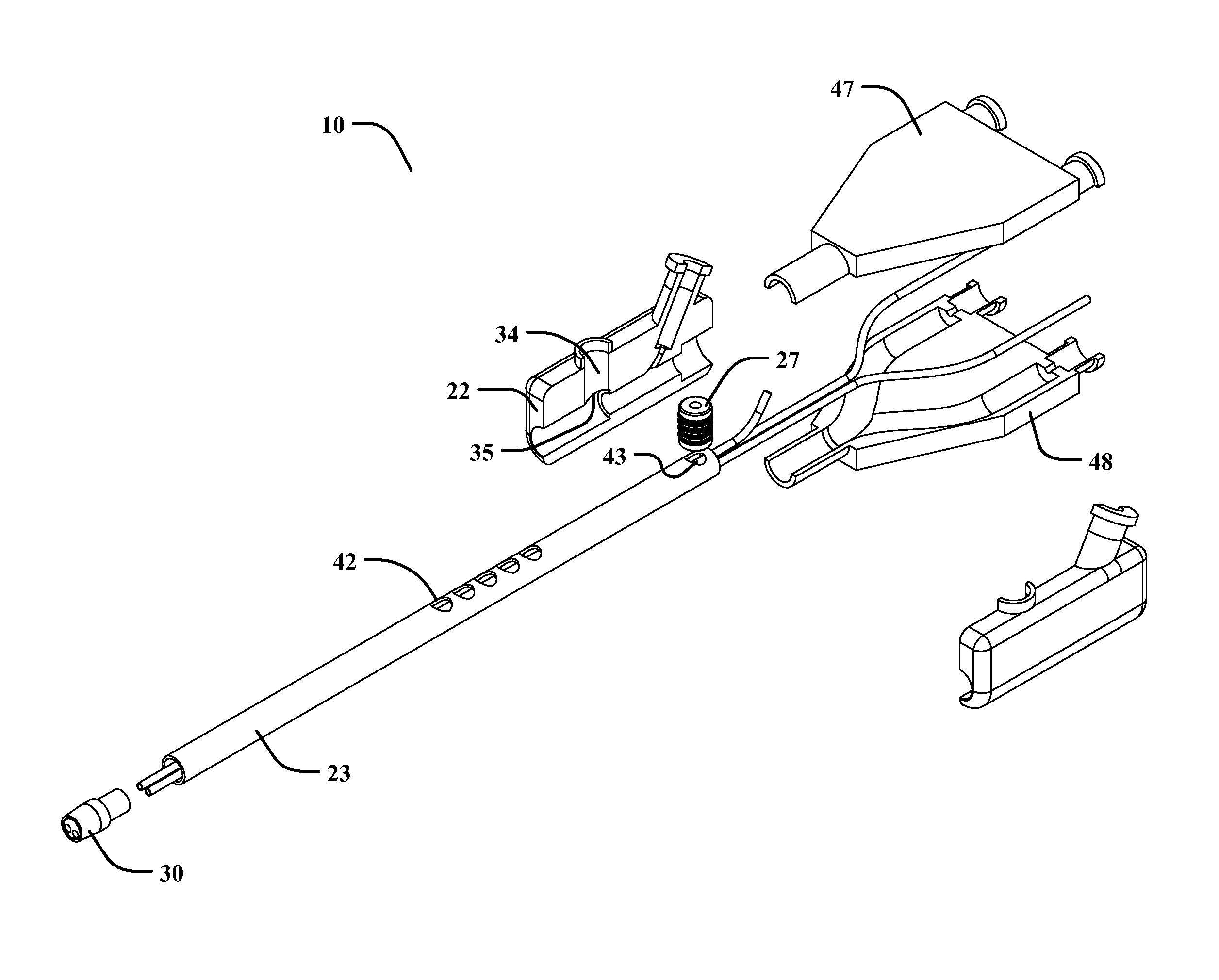
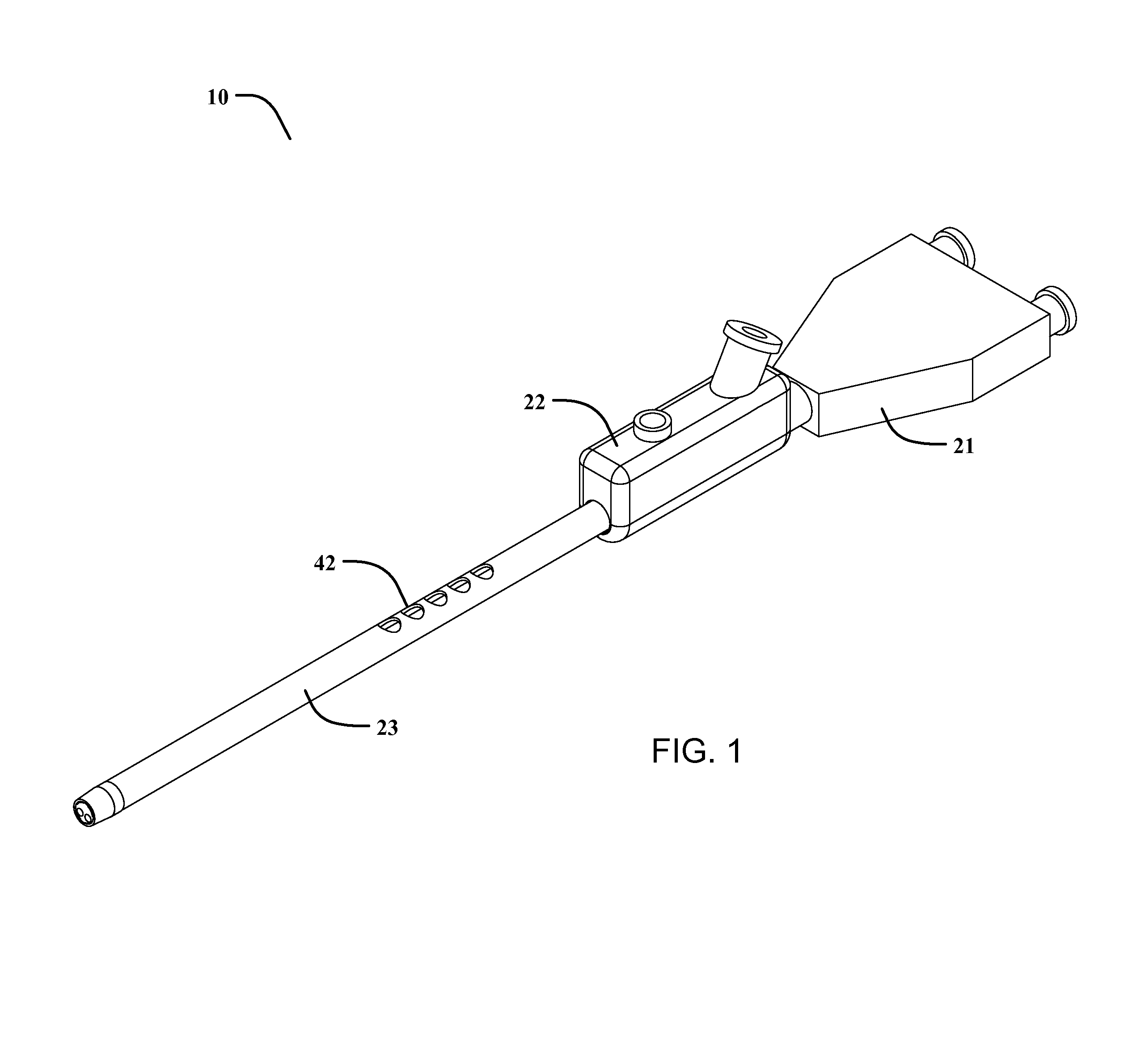
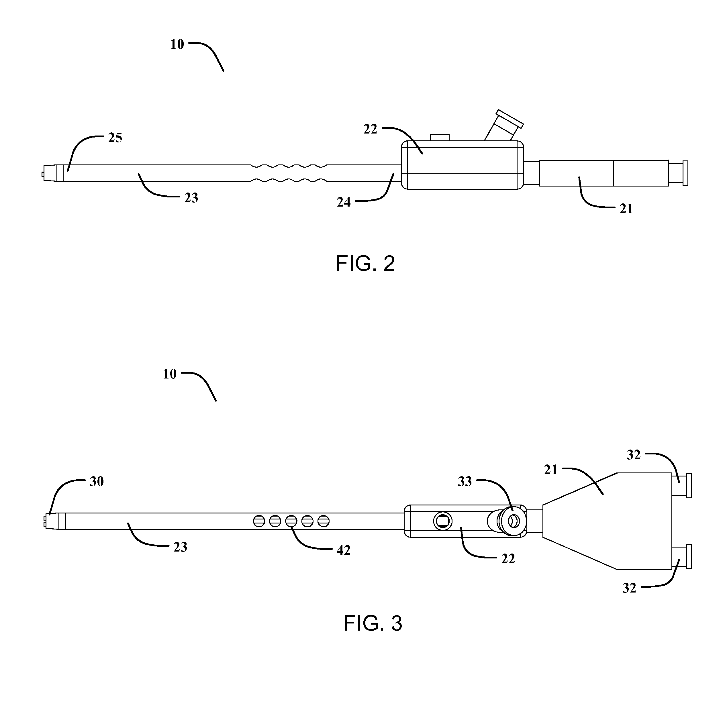
[0018]The present invention permits surgeons to introduce an endoscopic biomaterial applicator through a surgical trocar, creating an air tight seal, and then dispense one or a plurality of biomaterials into the surgical site. The surgeon will place the device into the trocar, where pressurized gas is channeled through the biomaterial applicator and through the spray tip for dispersing the biomaterial in a spray fashion.
[0019]The present invention will also utilize a gas release valve on the biomaterial applicator that is exposed to the internal pressure of the abdominal cavity, wherein the pressure within the abdominal cavity can be automatically released when spray actuation is initiated. Once spray actuation is initiated, the input gas will compress a control piston against a control spring, opening the gas release valve and venting the intra-abdominal pressure to the atmosphere. When spray actuation is terminated, the control spring will return the control piston to a closed position, preventing additional intra-abdominal pressure from venting to the atmosphere.

Problems solved by technology

Generally, surgical procedures produce a great deal of trauma for the human body, as large incisions are used to permit access to the internal tissue that requires attention.
Any pressures higher than this common pressure level can increase the risk of gas embolization to the patient.
However, even CO2 gas can be toxic to a human if high levels are absorbed into the blood stream.
With gas-assisted spray applicators that also introduce gas into the abdominal cavity in order to provide spray delivery of the biomaterial, this can pose problems for maintaining a safe intra-abdominal pressure.
If the flow rate of gas introduced by the biomaterial applicator is greater than the capacity of the insufflator to remove this sudden volume introduction of gas, the intra-abdominal pressure can rise above the desired intra-abdominal pressure setpoint, and subject the patient to potential gas embolism.
The first limitation of Hoogenakker is that it requires an independent gas regulator to provide the gas venting function required to reduce the risk of over-pressurization of the abdominal cavity.
This can pose cumbersome logistical issues in an operating room environment and also requires the coordination between two operating room personnel to make the tubing connections.
Furthermore, when objects are passed back and forth between the sterile side and non-sterile side of an operating room, additional risks related to infection can occur.
The second limitation of Hoogenakker is that the venting function of this system requires the available use of a gas vent port on a surgical trocar.
However, if other trocars are used in the procedure, these trocars commonly do not include a gas vent port to reduce the cost of the trocar.
Furthermore, for single incision laparoscopic procedures (SILS), only one trocar is used in the procedure and the single available gas port on the trocar must be used by the insufflator, providing no means to connect a biomaterial spray applicator venting feature that requires this trocar gas port to effectively perform its venting function.
However, the same limitation exist for Redl as they do for Hoogenakker, where an available trocar gas valve port may not be available on a trocar if only one trocar with a gas valve port is used and the remaining trocars do not include a gas valve port.

Method used


Image

  • Method and apparatus for a self-venting endoscopic biomaterial applicator
  • Method and apparatus for a self-venting endoscopic biomaterial applicator
  • Method and apparatus for a self-venting endoscopic biomaterial applicator

Examples


Embodiment Construction

[0036]Illustrative embodiments of the invention are described below. The following explanation provides specific details for a thorough understanding of and enabling description for these embodiments. One skilled in the art will understand that the invention may be practiced without such details. In other instances, well-known structures and functions have not been shown or described in detail to avoid unnecessarily obscuring the description of the embodiments.

[0037]Unless the context clearly requires otherwise, throughout the description and the claims, the words “comprise,”“comprising,” and the like are to be construed in an inclusive sense as opposed to an exclusive or exhaustive sense; that is to say, in the sense of “including, but not limited to.” Words using the singular or plural number also include the plural or singular number respectively. Additionally, the words “herein,”“above,”“below” and words of similar import, when used in this application, shall refer to this appli...


PUM


Abstract

The present invention will provide an endoscopic biomaterial applicator used during laparoscopic surgical procedures that utilizes pressurized gas to facilitate the spray application of a biomaterial while safely and automatically venting the additional volume of gas introduced into the abdominal cavity by endoscopic biomaterial applicator. This is accomplished utilizing a manifold, a plurality of channels, an elongated housing, a spray tip, a cannula gasket, and a gas release valve.

Description

CROSS-REFERENCE TO RELATED APPLICATIONS[0001]This application claims the benefit of U.S. Provisional Patent Application No. 61 / 667,906, filed on Jul. 3, 2012, and incorporated herein by reference.STATEMENT REGARDING FEDERALLY SPONSORED RESEARCH AND DEVELOPMENT[0002]Not Applicable.FIELD OF THE INVENTION[0003]This invention relates to an endoscopic biomaterial applicator, and more particularly, to an applicator that can self-vent gas introduced by the endoscopic applicator while providing a spray application of a biomaterial.DISCUSSION OF RELATED ART[0004]Generally, surgical procedures produce a great deal of trauma for the human body, as large incisions are used to permit access to the internal tissue that requires attention. Modern advancements in surgical procedures have given rise to minimally invasive surgeries, where small incisions are used and the stress on the human body is reduced. One popular type of minimally invasive surgery is laparoscopic surgery, where operations on th...

Claims


Application Information

Patent Timeline
Patent Type & Authority Applications(United States)
IPC IPC(8): A61M11/02
CPCA61M11/02A61M13/00
Inventor LYNCH, PATRICK
Owner LYNCH PATRICK
Why Eureka
Try Eureka