Minimally invasive methods for implanting a sacral stimulation lead

a sacral stimulation and minimally invasive technology, applied in the direction of spinal electrodes, internal electrodes, therapy, etc., can solve the problems of compromising urinary tract performance, affecting the function of urinary bladder, and affecting the urinary bladder

Active Publication Date: 2014-01-30
CIRTEC MEDICAL CORP
View PDF2 Cites 7 Cited by
  • Summary
  • Abstract
  • Description
  • Claims
  • Application Information

AI Technical Summary

Problems solved by technology

Many people suffer from an inability to control urinary function, i.e., urinary incontinence.
A variety of disorders may compromise urinary tract performance and contribute to incontinence.
For example, aging can often result in weakened sphincter muscles, which cause incontinence, or weakened bladder muscles, which prevent complete emptying.
Some patients also may suffer from nerve disorders that prevent proper triggering and operation of the bladder or sphincter muscles.
Fecal incontinence is the inability to control bowel function.
This takes time and creates extra trauma around the stimulation lead.

Method used


Image

  • Minimally invasive methods for implanting a sacral stimulation lead
  • Minimally invasive methods for implanting a sacral stimulation lead
  • Minimally invasive methods for implanting a sacral stimulation lead

Examples


Embodiment Construction

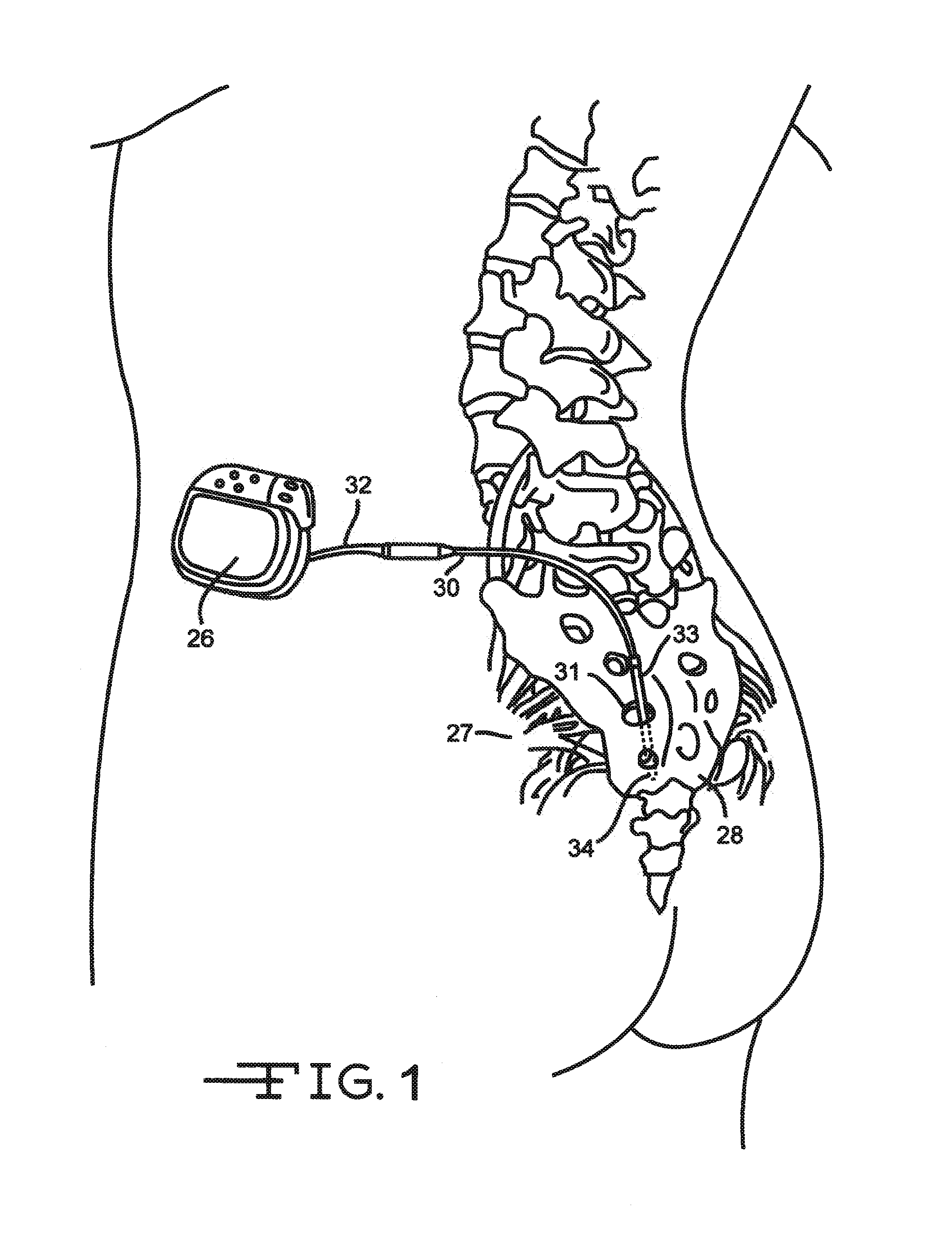
[0034]FIG. 1 shows an embodiment of an implanted neurostimulator 26 for stimulating sacral nerves 27 located near the sacrum 28. The sacral nerves are accessible through an entry point, in the skin along an insertion path 33 into a foramen 31 to reach a desired location 35. A neurostimulation system can include a stimulation lead 30, an optional lead extension 32, an implantable neurostimulator 26, a physician programmer (not shown), and a patient programmer (not shown). The stimulation lead 30 has electrical contacts 34 positioned on the distal end to stimulate nerves, and connectors (not shown) on the proximal end to connect to a lead extension or directly to the implantable neurostimulator 26.

[0035]The implantable neurostimulator 26 provides a programmable stimulation signal that is delivered to a desired location to stimulate selected nerves. The implantable neurostimulator 26 is typically implanted in a subcutaneous pocket around the upper buttocks, sometime after the stimulati...


PUM


Abstract

Methods and apparatus for implanting a neural stimulation lead in a patient's body are described. A lead assembly comprises a pointed-tip stylet, a stimulation lead, and an optional tube to deploy a fixation element attached to the lead. One embodiment of the implant methods starts with inserting the pointed-tip lead assembly directly into tissue. After the desired implant position is determined, the pointed-tip component is separated from the stimulation lead and removed from the tissue, leaving the stimulation lead implanted. After confirmation that the stimulation lead is in the right, tissue location, the pointed-tip component is removed from the body, leaving the stimulation lead in place. The stimulation lead can be connected to a neurostimulator to delivery therapies to treat neural disorders, such as urinary control disorders, fecal control disorders, sexual, dysfunction, and pelvic pain, etc.

Description

CROSS-REFERENCE TO RELATED APPLICATIONS[0001]This application is a divisional of U.S. patent application Ser. No 12 / 506,282, filed on Jul. 21, 2009, which claims priority from U.S. Provisional Application Ser. No. 61 / 082,271, filed Jul. 21, 2008.TECHNICAL FIELD[0002]The present invention relates to a neural stimulation lead assembly and minimally invasive implant methods associated with use of the described lead assembly. More specifically, the present invention relates to a lead assembly including a stimulation lead and a pointed-tip stylet received in a lumen of the lead. Minimally invasive method involves inserting the lead assembly into tissue without using a dilator.BACKGROUND OF THE INVENTION[0003]Many people suffer from an inability to control urinary function, i.e., urinary incontinence. Different muscles, nerves, organs and conduits within the urinary tract cooperate to collect, store and release urine. A variety of disorders may compromise urinary tract performance and con...

Claims


Application Information

Patent Timeline
Patent Type & Authority Applications(United States)
IPC IPC(8): A61N1/05
CPCA61N1/0558A61N1/0551A61N1/36057A61N1/00
Inventor YE, QINGSHANSWOYER, JOHN M.
Owner CIRTEC MEDICAL CORP
Why Eureka
Try Eureka