

Use of radiographic contrast agents for detecting dental caries
a radiographic contrast agent and caries technology, applied in the field of new imaging methods of oral and dental tissue, can solve the problems of inability to accurately distinguish between dental caries and the shape, inability of visual inspection and x-ray imaging to distinguish between cavitated and non-cavitated dental carious lesions between posterior teeth, and inability to infect the inflammation of the apical tissu
- Summary
- Abstract
- Description
- Claims
- Application Information
AI Technical Summary
Benefits of technology
Problems solved by technology
Method used
Image

Examples
example 1
Absorption of Topical Intra-Oral Composition Compared to Control
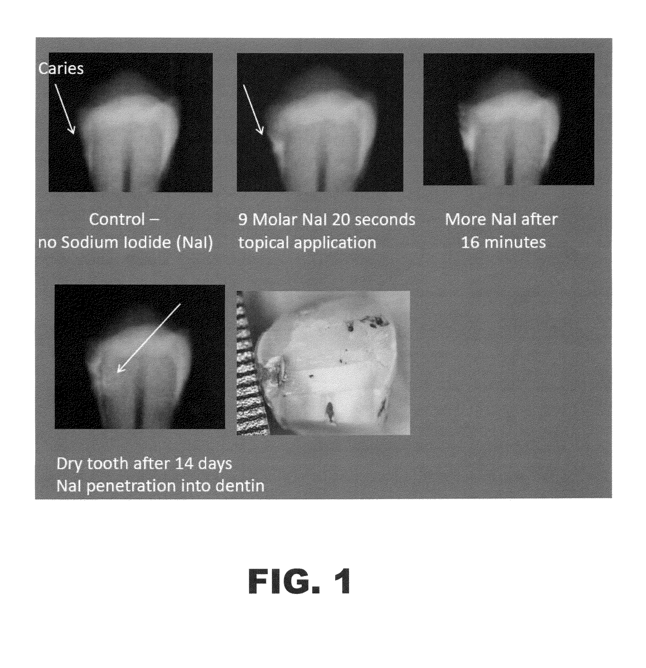
[0061]A radiographic contrast imaging analysis using x-ray imaging was performed, comparing the imaging of identified dental caries before and after administration of a topical intra-oral composition comprising sodium iodide and distilled water according to the methods of the current invention. Specifically, an x-ray image of a tooth with identified dental caries was performed, without administering the topical intra-oral composition. This image was used as a control for determining the ability of typical x-ray images to detect dental caries in a patient. Subsequently, the same tooth was administered a composition comprising sodium iodide and distilled water, with the composition having a sodium iodide concentration of 9 molar. After administration of the composition to the tooth, x-ray images were obtained 20 seconds after administration and 16 minutes after administration. Further, an x-ray image of the tooth was take...
example 2
Comparison of Absorption and Imaging Between Cavitated Surfaces and Sound Healthy Surfaces
[0063]An experiment was performed to test the ability of the topical intra-oral compositions and methods to differentiate between cavitated surfaces (dental caries) and healthy, sound surfaces. Specifically, an x-ray image of a tooth with an identified cavitated surface on one side of the tooth and a sound surface on the other side was performed, without administering the topical intra-oral composition. This image was used as a control for determining the ability of the topical intra-oral compositions to selectively absorb into cavitated surfaces, rather than sound surfaces. Subsequently, the same tooth was administered a composition comprising sodium iodide and distilled water, with the composition having a sodium iodide concentration of 9 molar. After administration of the composition to the tooth, an x-ray image was obtained 2 minutes after administration. The results are illustrated in FIG....
example 3
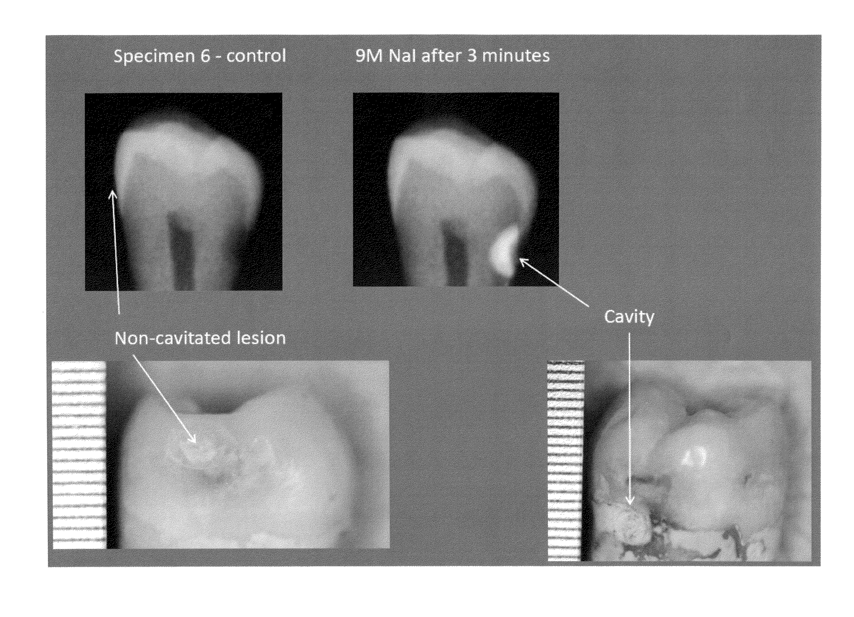
Comparison of Absorption and Imaging between Cavitated Lesions and Non-Cavitated Lesions
[0065]An experiment was performed to test the ability of the topical intra-oral compositions and methods to differentiate between cavitated surfaces (a cavity) requiring treatment and non-cavitated lesions not requiring treatment. Specifically, an x-ray image of a tooth with an identified cavitated surface on the right side of the tooth and a non-cavitated lesion on the other side was performed, without administering the topical intra-oral composition. This image was used as a control for determining the ability of the topical intra-oral compositions to selectively absorb into cavitated lesions, rather than non-cavitated lesions. Subsequently, the same tooth was administered a composition comprising sodium iodide and distilled water, with the composition having a sodium iodide concentration of 9 molar. After administration of the composition to the tooth, an x-ray image was obtained 3 minutes aft...
PUM
| Property | Measurement | Unit |
|---|---|---|
| concentration | aaaaa | aaaaa |
| concentration | aaaaa | aaaaa |
| concentration | aaaaa | aaaaa |
Abstract
Description
Claims
Application Information