Composition for treating intraventricular hemorrhage in preterm infants comprising mesenchymal stem cells
a technology of mesenchymal stem cells and stem cells, which is applied in the direction of skeletal/connective tissue cells, extracellular fluid disorders, peptide/protein ingredients, etc., can solve the problems that no histopathological abnormalities have been found in any organ including the lung of fully grown rats, and achieve the effect of preventing ventricular dilatation, reducing the level of inflammatory cytokines, and effectively preventing hydrocephalus
- Summary
- Abstract
- Description
- Claims
- Application Information
AI Technical Summary
Benefits of technology
Problems solved by technology
Method used
Image
Examples
example 1
Preparation of Intravascular Hemorrhage Model
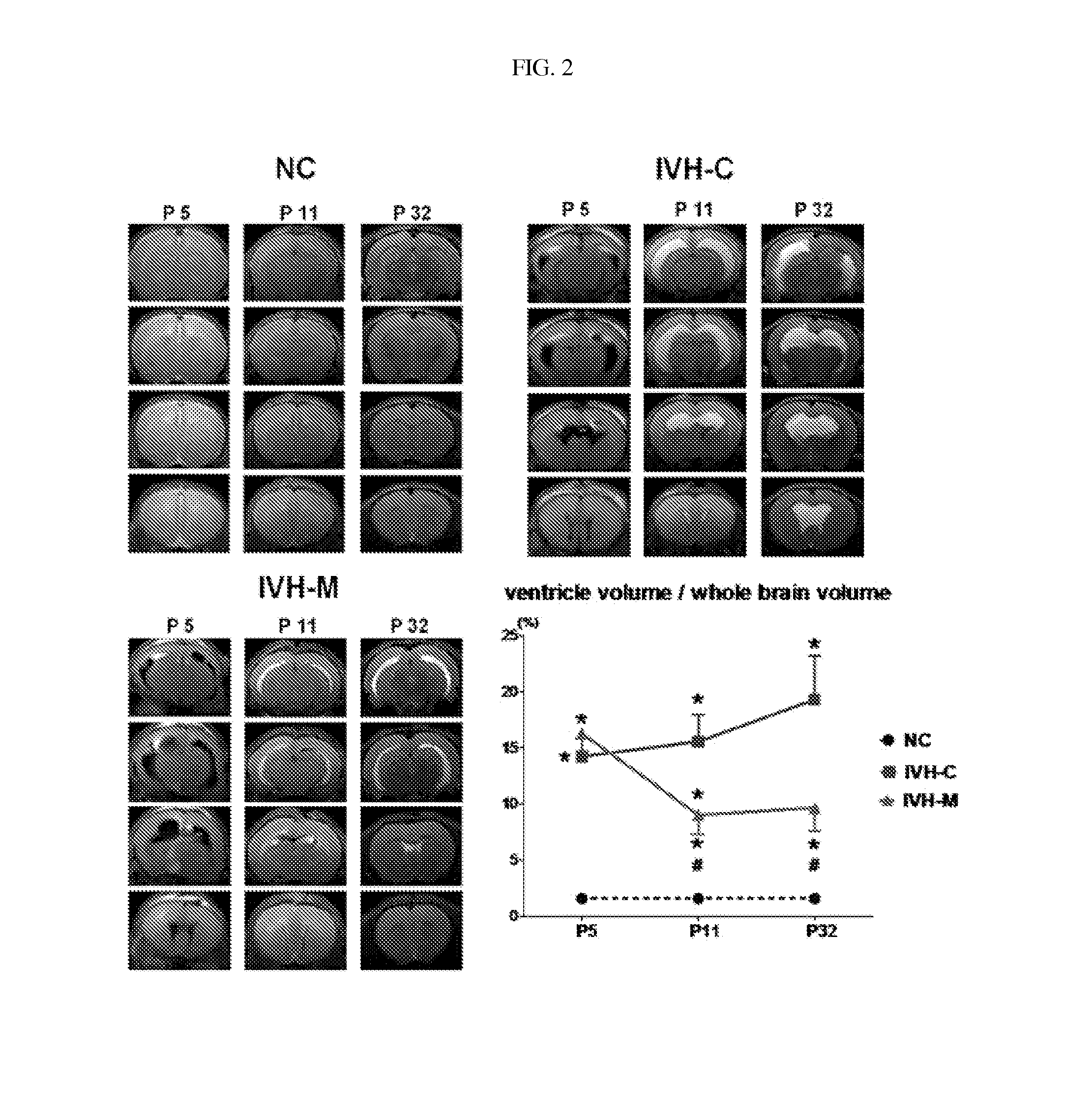
[0047]To establish intravascular hemorrhage models, blood was taken in a total volume of 200 μL from the tail vein of a mother and injected at a dose of 100 μL / ventricle into the left and the right ventricle of 4 days-old rats anesthetized with nitrogen monoxide (NO) slowly over 5 min using a 31 gauge syringe. At one day post-injection, the brains of the 5-days old rats were scanned by 7-teslar MRI to examine intravascular hemorrhage.
[0048]The brain MRI images were subjected to volumetric analysis using the Image J program to calculate the ventricular dilatation according to total ventricular volume / total brain volume. All of the intraventricular hemorrhage-induced rats were found to exhibit significant ventricular dilatation due to the accumulation of a significant amount of blood in the ventricles, compared to normal rats (NC), as measured by MRI at 5 days post-birth.
example 2
Administration of Umbilical Cord Blood-Derived Mesenchymal Stem Cells into Intraventricular Hemorrhage Model
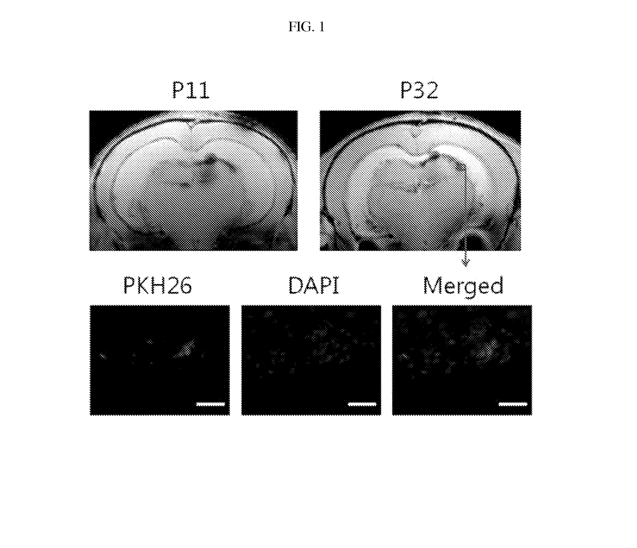
[0049]At 6 days post-birth, mesenchymal stem cells isolated from human umbilical cord blood were suspended in an amount of 1×10⁵ cells in 10 μL of PBS and slowly injected into the right ventricle of the neonatal rats anesthetized with NO gas.
[0050]At 11 days post-birth, the brains of all the neonatal rats were scanned by 7-teslar MRI (T2, Flash) to monitor ventricular dilatation.
[0051]At 32 days post-birth, final brain MRI (T2, Flash) was performed to trace the change of ventricular dilatation.
example 3
Preparation of Tissue Section
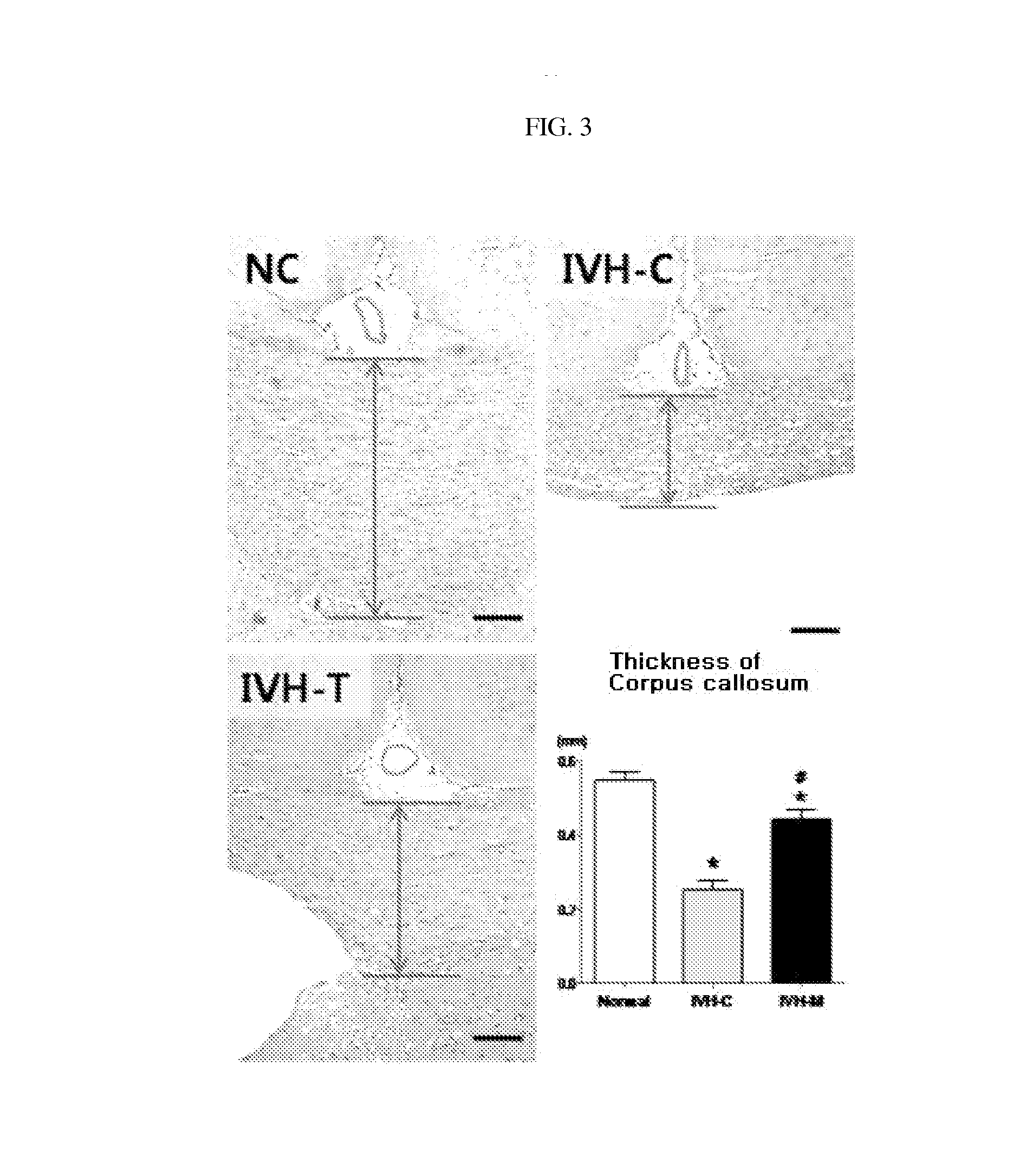
[0052]After the final MRI imaging, the neonatal rats were anesthetized by intraperitoneal injection of ketamine, and CSF was collected from the cisterna magna by cisternal tap with a 31 gauge syringe and frozen at −70° C. until use. The limbs were fixed before the rats were thoracotomized to expose the heart and the lungs. After a 23 gauge needle was fixed into the left ventricle, the right atrium was punctured and perfused with 4% paraformaldehyde. Then, an incision was made in the skull, and brain tissue was carefully taken and fixed in 4% formalin, or regions around the cerebral ventricles were sectioned, rapidly frozen in nitrogen gas, and stored at −70° C. until use.
PUM
| Property | Measurement | Unit |
|---|---|---|
| Cell death | aaaaa | aaaaa |
Abstract
Description
Claims
Application Information
 - R&D
- Intellectual Property
- Life Sciences
- Materials
- Tech Scout
- Unparalleled Data Quality
- Higher Quality Content
- 60% Fewer Hallucinations
Browse by: Latest US Patents, China's latest patents, Technical Efficacy Thesaurus, Application Domain, Technology Topic, Popular Technical Reports.
© 2025 PatSnap. All rights reserved.Legal|Privacy policy|Modern Slavery Act Transparency Statement|Sitemap|About US| Contact US: help@patsnap.com


