
Device, method and kit for the detection of different markers in different cellular or molecular types and their quantifications

a technology of different cellular or molecular types and detection methods, applied in the field of devices, methods and kits for the detection of different markers in different cellular or molecular types and their quantification, can solve problems such as incompatibility with the arrangement of rapid assays

Inactive Publication Date: 2014-04-24
MAZZEO ALESSANDRA
View PDF4 Cites 2 Cited by
  • Summary
  • Abstract
  • Description
  • Claims
  • Application Information

AI Technical Summary

Benefits of technology

The patent text describes a solution for diagnosing and treating tuberumberculosis in humans and animals. The solution includes a kit that is easy to use and can be performed by veterinary staff with little training and equipment. This kit helps identify infected animals at an early stage to prevent the spread of the infection in livestock farming. Overall, this solution helps improve the diagnosis and treatment of tuberugoa infections in both humans and animals.

Problems solved by technology

Differentiating the bovine active tuberculosis from the latent one is irrelevant, since the national and international compulsory eradication programmes do not allow the infected cattle to be raised, at any infection stage.
In the state of the art, there are no systems for the arrangement of rapid diagnostic kits detecting the host cellular immune response to pathogenic microorganisms causing infections not detectable by antibody assays; moreover, equipment-free systems—based on the simultaneous detection of different cells exhibiting infection specific markers simultaneously quantifiable—are not reported; additionally, there are not solid phases for immumoenzymatic tests and ELISA useful for performing assays aimed to the simultaneous detection of both different markers and the different cells that exhibit them.
Moreover, the cell types could be identified carrying out further tests in the recovered liquid; this is incompatible with the arrangement of rapid assays.
U.S. 2010 / 083778 describes devices suited for retaining nonporous substrates which are simultaneously screened; they are absolutely unadapt for arranging the kits presented here, since they do not provide the separation into individual wells, which is essential for the correct diagnostic marker quantification, as illustrated here.

Method used


Image

  • Device, method and kit for the detection of different markers in different cellular or molecular types and their quantifications
  • Device, method and kit for the detection of different markers in different cellular or molecular types and their quantifications
  • Device, method and kit for the detection of different markers in different cellular or molecular types and their quantifications

Examples


Embodiment Construction

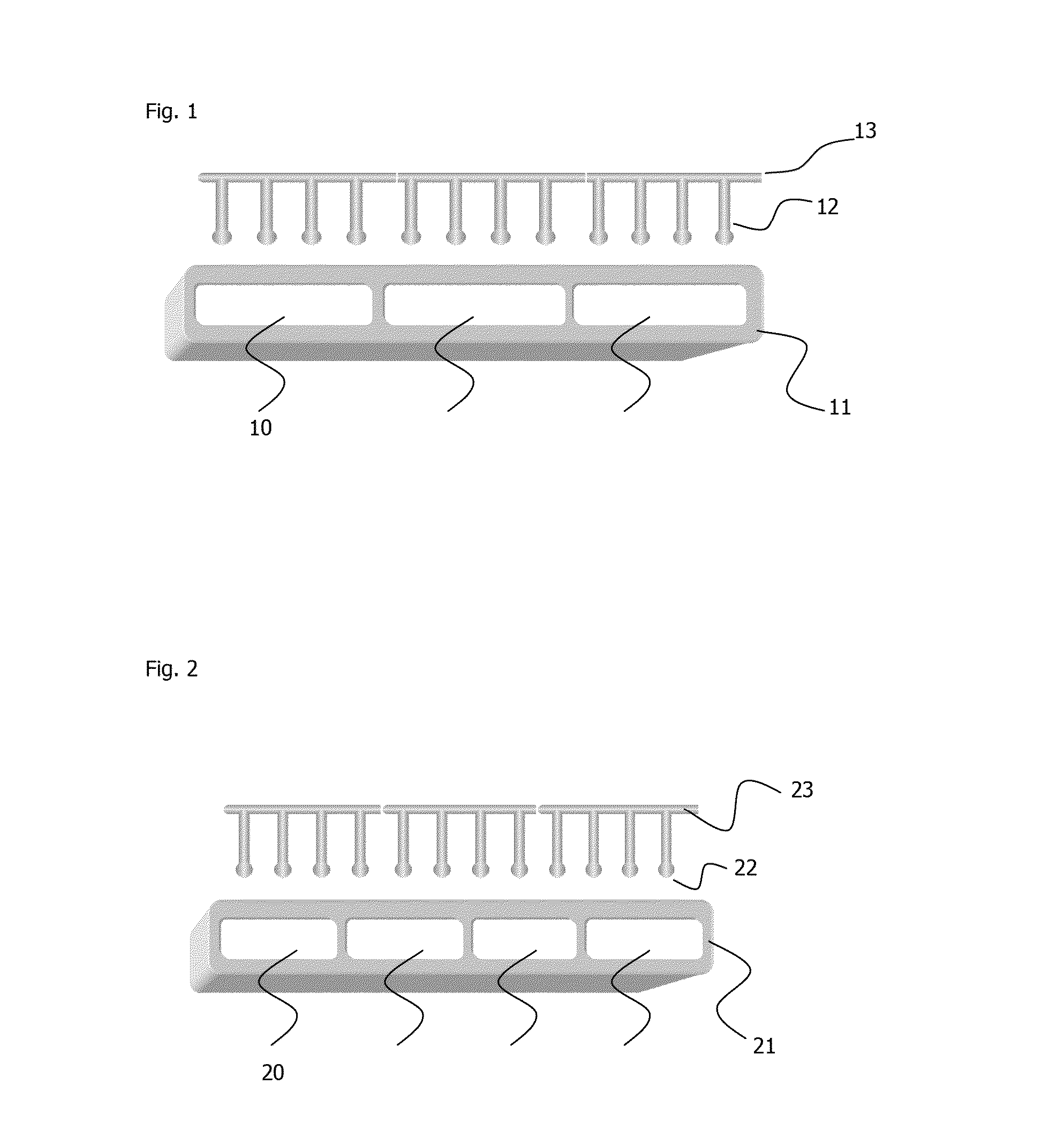
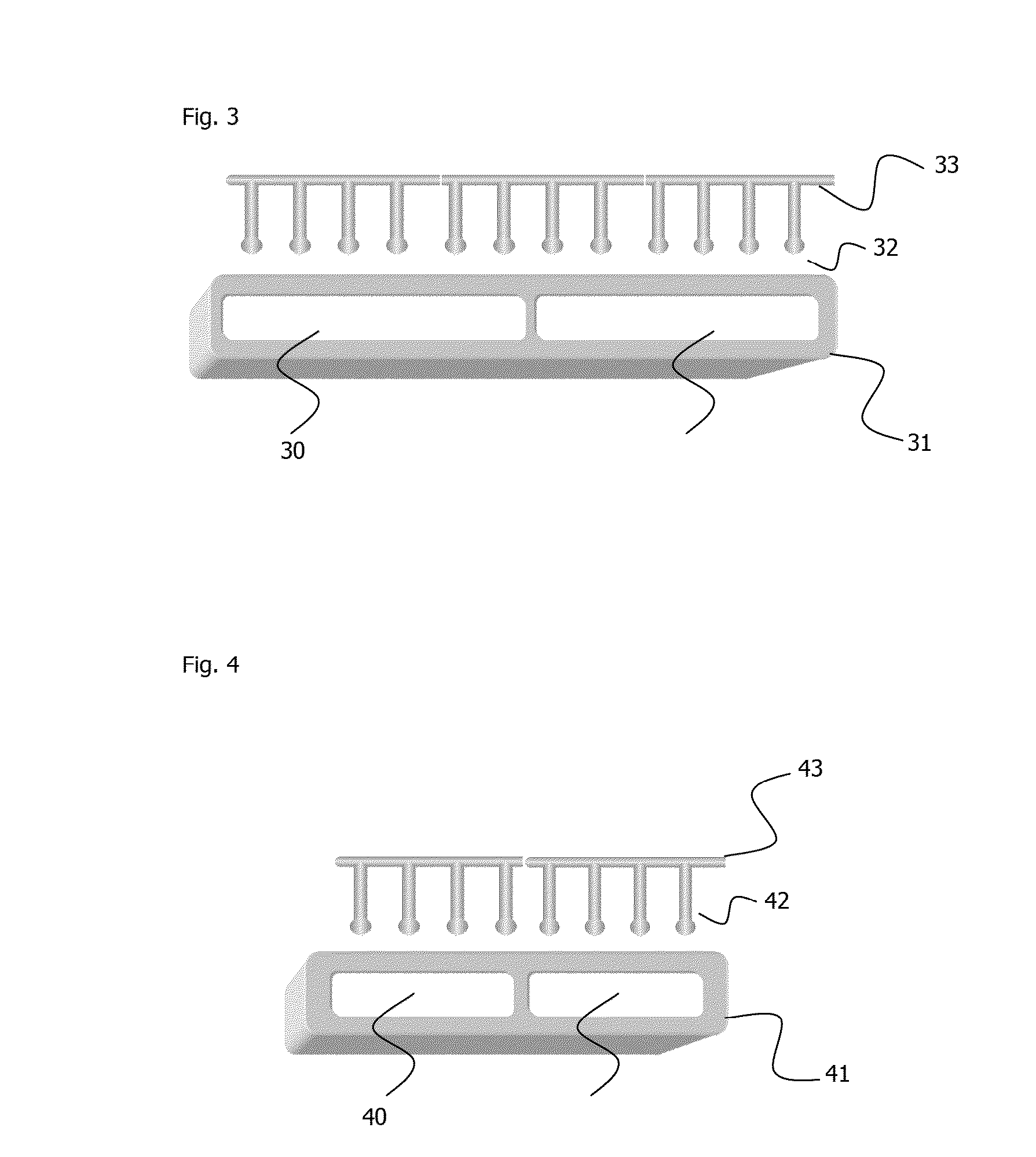
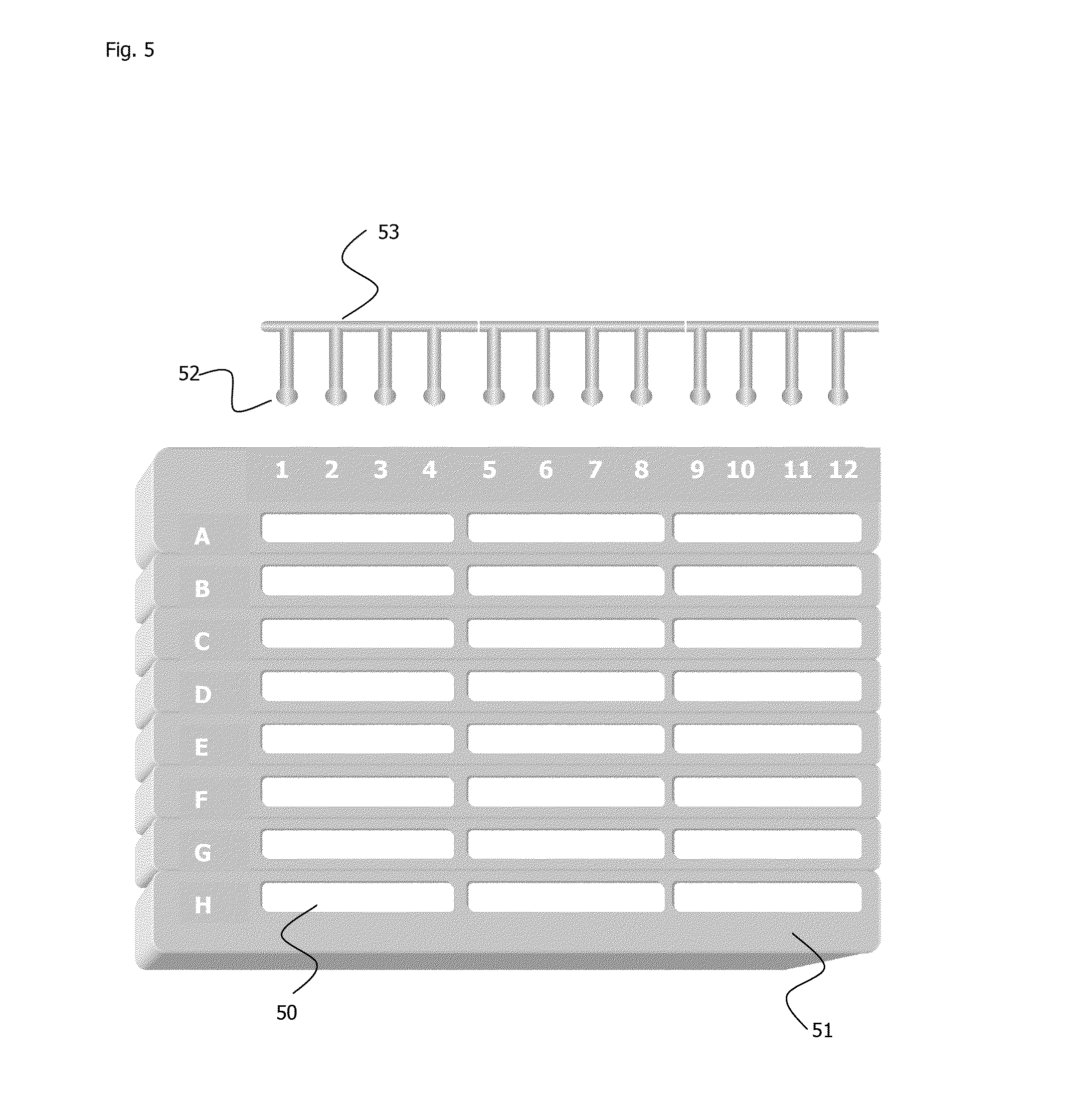
[0025]The present invention concerns:[0026]devices in the shape of microplates or microstrips having wells with extended length apt to receive 3 or 4 or 6 immunosorbent elements protruding from a rod at the same modular distance of wells arranged in standard 8-wells microstrips or in 12-wells microstrips or in standard 96-wells microplates;[0027]the method that uses these devices making two different shaped solid phases compete with each other; a solid phase is constituted by the extended wells, each of which immobilize a different cell or molecule in the examined sample; the other solid phase is constituted by immunosorbent elements protruding from a rod, each of which has been previously coated with one of the same markers to be detected in the sample; then, these immunosorbent elements are dipped into each extended well in groups of 3 or 4 or 6, after ligands for markers to be detected have been added in liquid phase in the extended wells; these ligands bind to the immunosorbent ...


PUM


Abstract

Microplates or microstrips having wells with extended length to receive immunosorbent elements protruding from a rod at the same modular distance of wells arranged in standard microstrips or microplates and methods of use are provided. A solid phase is constituted by the extended wells, each of which immobilize a different type of cell or molecule in the sample; the other solid phase is constituted by immunosorbent elements protruding from a rod, each of which has been previously coated with one of the same markers to be detected in the sample. Ligands for markers to be detected are added in a liquid phase to the wells; allowing the ligands competitively bind to the immunosorbent elements, inversely proportional to the quantity of the markers of each type of cell or molecule immobilized on the proper extended well. These ligands are simultaneously quantifiable by an immunoenzymatic assay using chromogenic substrate and a spectrophotometer.

Description

CROSS REFERENCE TO RELATED APPLICATION[0001]This application is a continuation of prior filed International Application No. PCT / IB2012 / 052021, as filed on Apr. 23, 2012, entitled “Device, Method And Kit For The Detection Of Different Markers In Different Types Of Cells Or Molecules And Their Quantification”, which claims priority to Italian Application No. CS2011A000012, as filed on Apr. 21, 2011, and Italian Application No. CS2012A000019, as filed on Apr. 20, 2012. The contents of which are herein incorporated by reference.BACKGROUND OF THE INVENTION[0002]The indirect diagnosis of tuberculosis, caused by Mycobacterium tuberculosis, one of the most worldwide spread infection among the human population, cannot be carried out by the standard serological tests and it is based on the cellular response detection methods, such as:[0003]in vivo skin test, that has to be read after 72 hours, which is useful to detect infection cases regardless of the mycobacterium localization, based on a I...

Claims


Application Information

Patent Timeline
IPC IPC(8): G01N33/569
CPCG01N33/5695B01L3/5085B01L2300/021B01L2300/0829G01N33/54366
Inventor MAZZEO, ALESSANDRA
Owner MAZZEO ALESSANDRA
Learn More