Electrophysiological monitoring of uterine contractions

a technology of uterine contractions and electrical monitoring, applied in the field of electrical monitoring of uterine contractions, can solve the problems of inability to discriminate between uterine contractions and (in)voluntary contractions of abdominal muscles, unreliable onset and amplitude of reflected activity, and inability to accurately detect the contractions. , to achieve the effect of reducing the risk of uterine contractions, and improving the quality

Active Publication Date: 2014-06-26
NEMO HEALTHCARE
View PDF4 Cites 31 Cited by
  • Summary
  • Abstract
  • Description
  • Claims
  • Application Information

AI Technical Summary

Benefits of technology

[0007]It is an object of the invention to provide a signal processing arrangement for and a signal processing method of processing electrophysiological signals related to uterine contractions which have a relatively short delay between the moment in time when electrophysiological signals related to uterine activity are measured and the moment in time when the processed uterine activity data becomes available to a user or to other devices which uses the processed uterine activity data as input data.

Problems solved by technology

With this external method however, it is not always possible to discriminate between uterine contractions and (in)voluntary contractions of the abdominal muscles (e.g. due to movement, coughing, etc.).
In addition, in case the tocodynamometer does detect true uterine contractions, both the onset and amplitude of the reflected activity may be unreliable and inaccurate.
However, due to the risks involved, e.g. the catheter has been reported to pierce the placenta, and the fact that the IUPC can only be applied after rupture of the membranes and sufficient dilatation of the cervix, the use of this method in current clinical practice is limited.
However, in all methods that have been proposed, such a delay exists either due to processing that requires a certain amount of data, or due to filter characteristics (such as in the above cited patent application).
As a result, these methods fail to provide an instantaneous output that can directly be used for cardiotocography.
Additionally, the electrical measure for uterine activity that is provided by some of the methods suffers from a lack of robustness.

Method used


Image

  • Electrophysiological monitoring of uterine contractions
  • Electrophysiological monitoring of uterine contractions
  • Electrophysiological monitoring of uterine contractions

Examples


Embodiment Construction

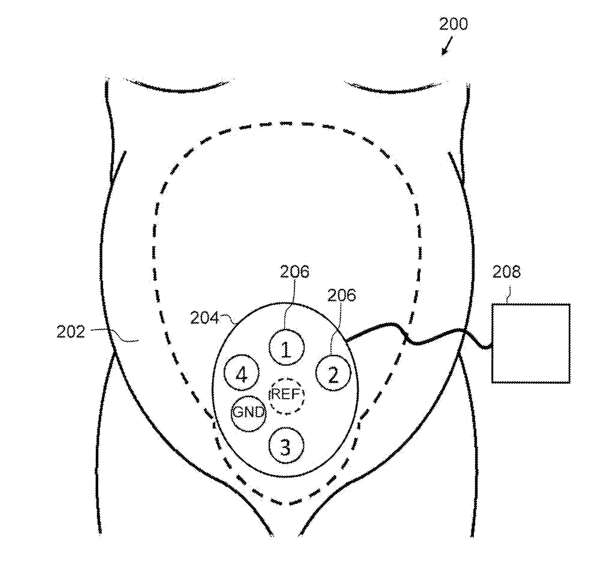
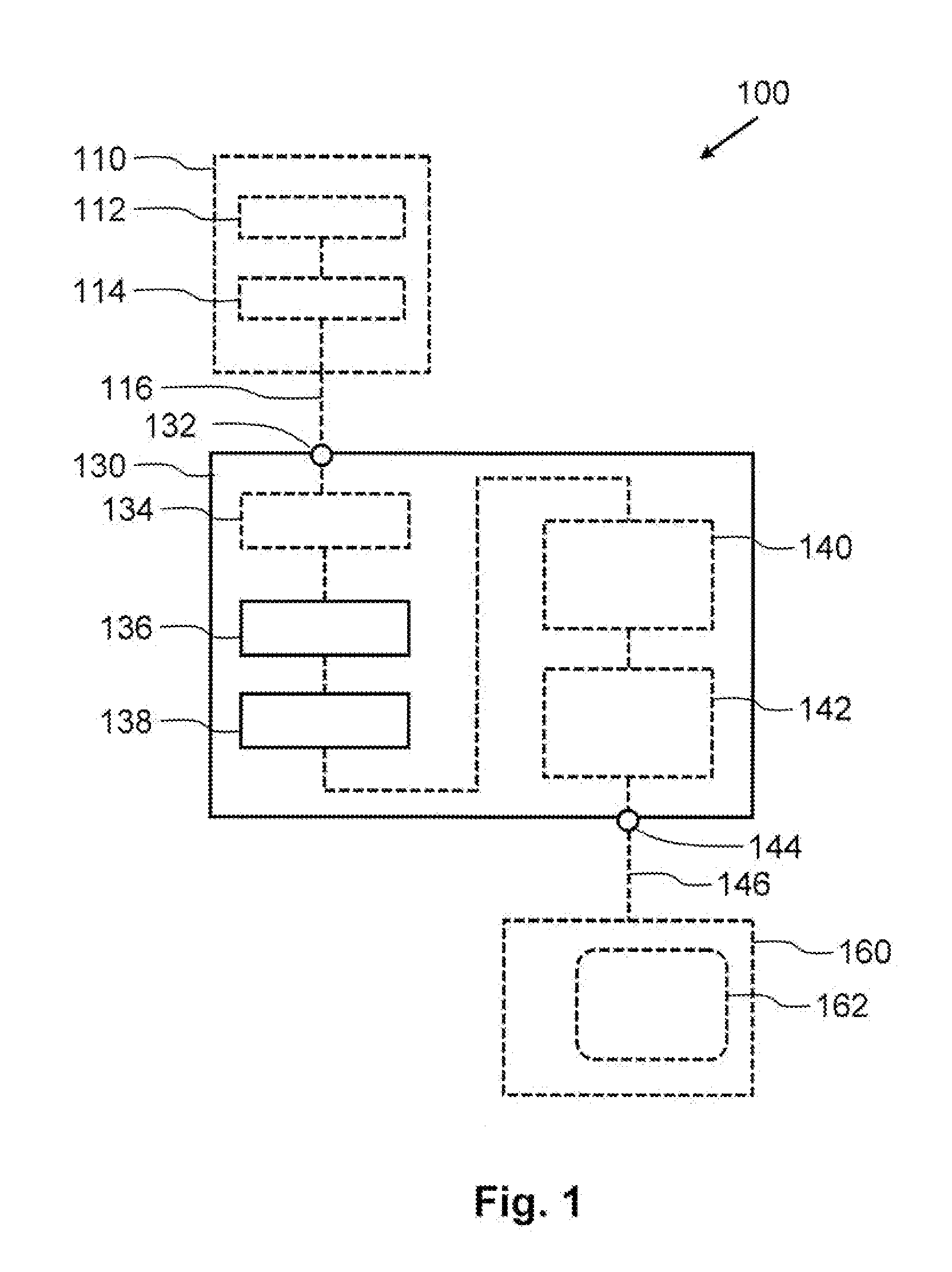
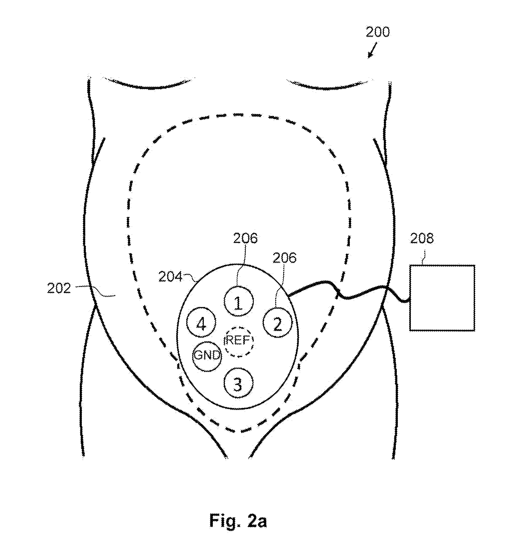
[0047]A first embodiment is shown in FIG. 1. FIG. 1 schematically shows a monitoring system 100 which comprises a physiological measurement system 110, a signal processing arrangement 130, and an optional presentation device 160.

[0048]The physiological measurement system 110 comprises at least two cutaneous or capacitive electrodes 112 which form a sensor for measuring activity of the muscles of the uterus of a pregnant woman. The at least two cutaneous or capacitive electrodes 112 are coupled to a physiological measurement circuit 114 which uses the two cutaneous or capacitive electrodes 112 to provide an electrophysiological signal 116. The physiological measurement circuit 114 at least amplifies the signal provided by the two cutaneous or capacitive electrodes 112 and may optionally digitalize the amplified signal. Different physiological measurement systems are known in the art and may be used in the monitoring system 100. The used physiological measurement system 110 must be su...


PUM


Abstract

A signal processing arrangement 130, a monitoring system 100, a signal processing method, a monitoring method of monitoring uterine contractions of a pregnant woman, and a computer program product are provided. The signal processing arrangement 130 receives an electrophysiological signal 116 representing uterine muscle activity of a pregnant woman at an input 132. A filter 136 generates a filtered electrohysterogram signal from the electrophysiological signal 116. The filter 136 allows the passage of spectral components between 0 and 3 Hz. A window function applicator 138 applies a window function to the filtered electrohysterogram signal to obtain an output waveform 146. The window function defines that samples of a time interval preceding the application of the window function need to be used The output waveform 146 simulates output data of tocodynamometer or an intra-uterine pressure catheter. The output waveform 146 is provided at an output 144 of the signal processing arrangement.

Description

FIELD OF THE INVENTION[0001]The invention relates to a device for and a method of monitoring uterine contractions.BACKGROUND OF THE INVENTION[0002]For more than three decades, the worldwide standard for monitoring fetal condition has been cardiotocography (CTG). Cardiotocography is the simultaneous registration of fetal heart rate and uterine activity. In case of uterine contractions, the umbilical cord may get (partially) occluded. In addition, pressure might be exerted on the fetal head. A healthy fetus generally will respond to these new circumstances by a sudden reduction in fetal heart rate. Whether the fetal heart rate is indeed reduced and also the latency between the actual contraction and the deceleration of fetal heart rate, are important clinical parameters to diagnose fetal wellbeing. Two methods are well-known for monitoring the uterine contractions. The first of these methods consists of an elastic belt that contains a strain gauge to assess the degree of “hardness” of...

Claims


Application Information

Patent Timeline
Patent Type & Authority Applications(United States)
IPC IPC(8): A61B5/0488A61B5/00
CPCA61B5/4356A61B5/725A61B5/391A61B5/7203
Inventor PETERS, CHRISTIAAN HENDRIK LEONARDVULLINGS, RIK
Owner NEMO HEALTHCARE
Why Eureka
Try Eureka