Method for Calibrating a Counting Digital X-Ray Detector, X-Ray System for Performing Such a Method and Method for Acquiring an X-Ray Image

a digital x-ray detector and counting technology, applied in the field of calibration of counting digital x-ray detectors, can solve the problems of detectors with adjustable threshold values of discriminators, inability to continue counting without error, and noise of threshold values, and achieve the effect of high image quality

Active Publication Date: 2014-07-03
SIEMENS HEALTHCARE GMBH
View PDF1 Cites 24 Cited by
  • Summary
  • Abstract
  • Description
  • Claims
  • Application Information

AI Technical Summary

Benefits of technology

[0015]The count rate of the pixel elements is therefore measured, for example, in the case of X-ray irradiation with an X-ray spectrum of a commercially available X-ray source (e.g., max. 50, 70, 90 or 120 keV), while a first threshold value that is the same for all pixel elements is applied, for example, using discriminators and digital-to-analog converters. A further measurement is subsequently carried out at a further threshold value that is different from the first threshold value. It is of advantage if at least three or more measurements are performed at other threshold values. A plurality of measurements may also be carried out. For example, a series of different threshold values may be applied in ascending or descending order, and corresponding measurements may be taken. The measurement results are stored and used accordingly for determining and / or calculating individual correction threshold values that are to be used for planned measurements with the X-ray detector in addition to the originally identical threshold values for all of the pixel elements. The correction threshold values are selected such that for the defined X-ray spectrum that is the same for all pixel elements, all the pixel elements start with a count at essentially the same X-ray energy or differ from one another by only a small amount at least with respect to the start of the count.
[0016]A significant improvement in image quality during X-ray imaging and, for example, a reduction in threshold-value-induced noise may be achieved by the method according to one or more of the present embodiments. The method is a robust method that may also be reliably performed without monoenergetic (e.g., radioactive) X-ray sources, simply using typical X-ray spectra. As a result, the method may be performed without problems with any X-ray source. The method may easily be automated, thus enabling a recalibration to be performed quickly and reliably (e.g., in the case of drifts, temperature influences, etc).
[0020]According to an embodiment, the characteristic curves and / or the average value curves are fitted and / or smoothed and / or scaled. In this way, it is easier to determine or calculate the correction threshold values, statistical variations may be reduced, and the calibration may be improved. It may, for example, be provided initially to smooth the characteristic curves and adapt a suitable fit function to the individual characteristic curves. An average value curve is determined, for example, from the smoothed and fitted functions. The horizontal differences are likewise calculated, for example, based on the smoothed and fitted functions.
[0025]Initially, correction threshold values determined for the respective X-ray spectrum used are now present. In order to make the calibration more comprehensively applicable to a plurality of or even to all possible X-ray spectra and X-ray energies, the following acts are advantageously performed: the performing of the threshold values scan and the storing of the count rates are performed for at least two X-ray spectra, characteristic lines are generated from the measurements for the at least two X-ray spectra (e.g., using a fit function), for the pixel elements by plotting the maximum energy of the X-ray spectra against the threshold value at which the respective pixel element starts to count, an average value curve is determined from the characteristic lines of the pixel elements, and the characteristic lines are used for determining or calculating individual correction threshold values for the individual pixel elements. Correction threshold values are determined for a plurality of X-ray energies. Using this link between the energy of the X-ray spectra and the threshold values, and advantageously also using a fit function that connects the discrete points of the actually measured X-ray spectra, the corresponding correction threshold values may be derived for each pixel element and for each desired X-ray spectrum or each X-ray energy from the characteristic lines and, for example, to input the values into the X-ray detector. Accordingly, the method is able, using normal X-ray sources, to generate an absolute relationship between threshold value and energy.
[0027]In one or more of the present embodiments, a method for acquiring at least one X-ray image using an X-ray system is provided. Correction threshold values determined by a method according to one or more of the present embodiments are used during the acquisition of X-ray images for the purpose of correcting the threshold value noise. Such a method achieves X-ray images that are almost or completely free of threshold value noise and therefore exhibit a particularly high image quality.

Problems solved by technology

This readout process requires a certain amount of time during which it is not possible to continue counting without error.
Counting detectors with adjustable discriminator threshold values exhibit problems such as the following.
However, since only X-ray quanta having X-ray energy that lies above the threshold values are counted, different portions of the energy spectrum are counted, resulting in “threshold value noise”.
Such effects may lead to structural or “fixed pattern”-like noise.

Method used


Image

  • Method for Calibrating a Counting Digital X-Ray Detector, X-Ray System for Performing Such a Method and Method for Acquiring an X-Ray Image
  • Method for Calibrating a Counting Digital X-Ray Detector, X-Ray System for Performing Such a Method and Method for Acquiring an X-Ray Image
  • Method for Calibrating a Counting Digital X-Ray Detector, X-Ray System for Performing Such a Method and Method for Acquiring an X-Ray Image

Examples


Embodiment Construction

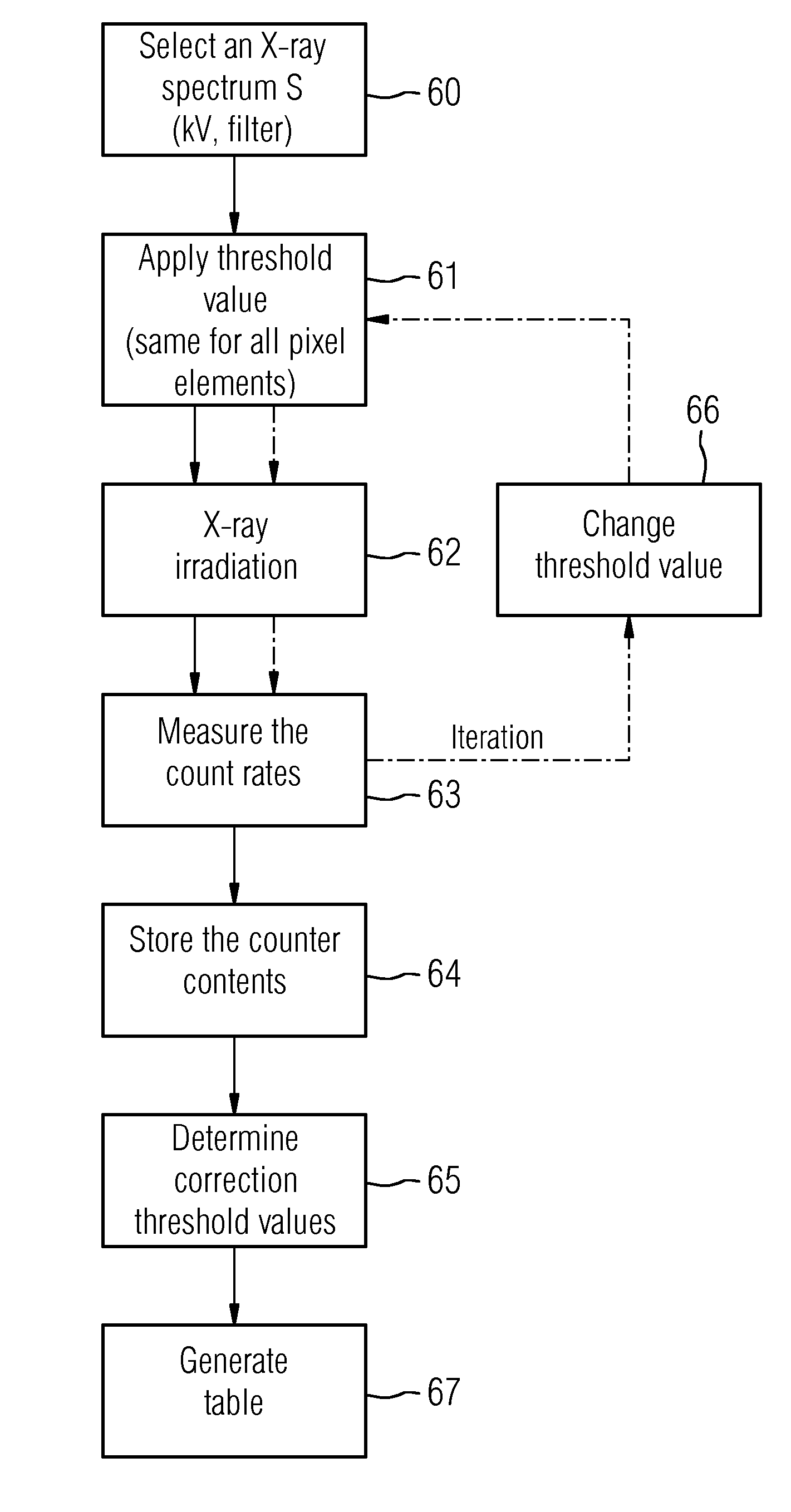
[0048]The present embodiments describe a method for calibrating a digital counting X-ray detector, by which a threshold value noise may be reduced or completely avoided. As a result, the quality of the X-ray imaging is significantly improved. An X-ray detector that may be calibrated by a method of the type has, for example, a structure such as described with reference to FIG. 3, including a direct converter 24 (e.g., CdTe or CZT) for converting X-ray quanta into electrical signals and a plurality of pixel elements in a matrix structure. The plurality of pixel elements may receive and register the signals as count events as a function of position provided the signals lie above a threshold value. The individual pixel elements have central functional elements, as shown, for example, in FIG. 5 (simply discriminating) or FIG. 7 (energy-discriminating). The threshold value that may be applied to the respective pixel element is adjustable.

[0049]Discriminators and analog-to-digital converte...


PUM


Abstract

A method for calibrating a counting digital X-ray detector includes performing a threshold value scan in at least one defined X-ray spectrum for irradiating the X-ray detector, which includes a matrix composed of pixel elements, storing count rates of the pixel elements as a function of respective applied threshold values, and from results of a measurement of count rates of the pixel elements, determining or calculating individual correction threshold values for the individual pixel elements. The individual correction threshold values correct a threshold value that is to be applied to the pixel elements for the defined X-ray spectrum such that threshold value noise is reduced.

Description

[0001]This application claims the benefit of DE 10 2013 200 021.9, filed on Jan. 2, 2013, which is hereby incorporated by reference in its entirety.BACKGROUND[0002]The present embodiments relate to calibrating a counting digital X-ray detector.[0003]X-ray systems are used for imaging for diagnostic examination purposes and for interventional procedures (e.g., in cardiology, radiology and surgery). X-ray systems 16, as shown in FIG. 1, have an X-ray tube 18 and an X-ray detector 17 (e.g., jointly mounted on a C-arm 19), a high-voltage generator for generating the tube voltage, an imaging system 21 (e.g., including at least one monitor 22), a system control unit 20 and a patient table 23. Biplane systems (e.g., having two C-arms) are likewise employed in interventional radiology. Generally, flat-panel X-ray detectors find application as X-ray detectors in many fields of medical X-ray diagnostics and intervention (e.g., in radiography, interventional radiology, cardioangiography), but ...

Claims


Application Information

Patent Timeline
Patent Type & Authority Applications(United States)
IPC IPC(8): G01T7/00
CPCG01T1/247G01T7/005
Inventor REITZ, SILKESPAHN, MARTIN
Owner SIEMENS HEALTHCARE GMBH
Why Eureka
Try Eureka