Method and optical system for evaluating concentrations of components in tissue

a tissue and optical system technology, applied in the field of tissue concentration evaluation, can solve the problems of scarring on the skin, participants may not accept the disadvantages aforementioned, long measurement time, and high system cost, and achieve the effect of non-invasiv

Active Publication Date: 2014-07-24
NAT CHENG KUNG UNIV
View PDF3 Cites 11 Cited by
  • Summary
  • Abstract
  • Description
  • Claims
  • Application Information

AI Technical Summary

Benefits of technology

[0012]The object of the present invention is to provide a method for evaluating the collagen concentration of tissues. In addition, other tissue chromophores, such as melanin, hemoglobin, and water can be extracted in the same time. More particularly, the method of the present invention can quantify the concentrations of the aforementioned components in a non-invasive way quickly and accurately.

Problems solved by technology

However, the slide examination is an invasive examination, and the wound for getting the slice may turn into scars on skin.
Hence, some participants may not accept the aforementioned disadvantages.
Although the multi-photon microscopy can provide structural information of tissues in vivo, this technique has disadvantages of long measurement time and high system cost.
However, the current detecting method still cannot accurately identify the differences between hypertrophic scars and keloids.

Method used


Image

  • Method and optical system for evaluating concentrations of components in tissue
  • Method and optical system for evaluating concentrations of components in tissue
  • Method and optical system for evaluating concentrations of components in tissue

Examples


Embodiment Construction

[0039]The present invention has been described in an illustrative manner, and it is to be understood that the terminology used is intended to be in the nature of description rather than of limitation. Many modifications and variations of the present invention are possible in light of the above teachings. Therefore, it is to be understood that within the scope of the appended claims, the invention may be practiced otherwise than as specifically described.

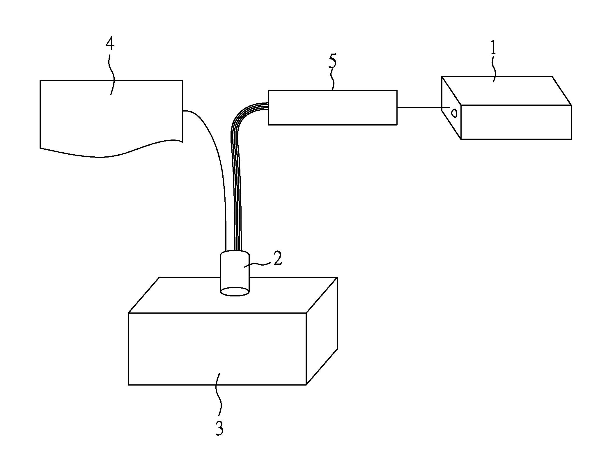
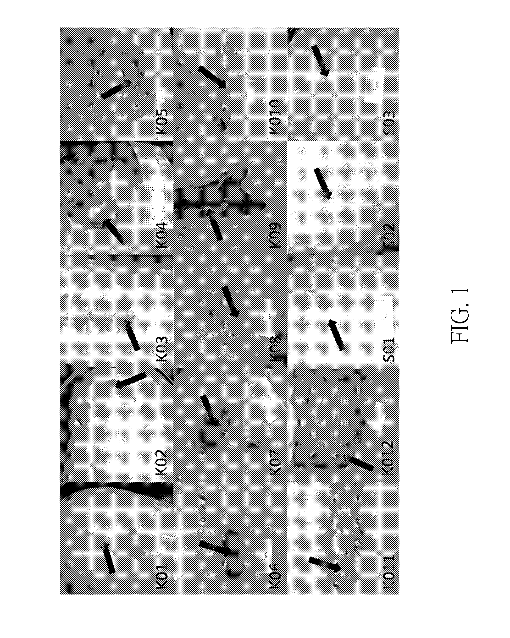
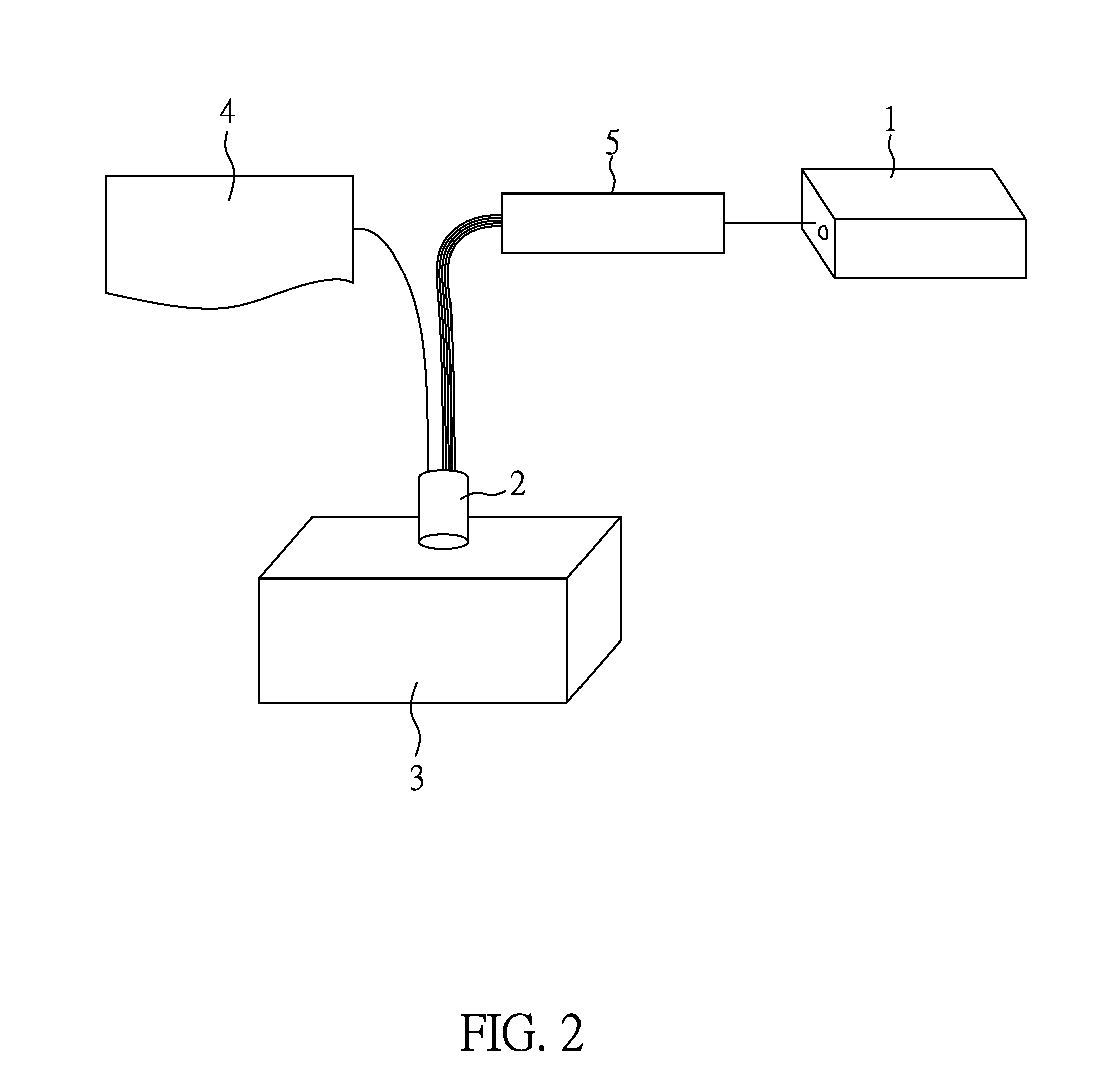
[0040]The following embodiments of the present invention demonstrated the detective accuracy of the method and the optical device of the present invention via quantifying the collagen contents at keloid sites and normal sites of the tested skin.

[0041]In the present embodiment, twelve subjects with keloid scars, and three subjects with normal scars were recruited in the National Cheng Kung University Hospital. The protocol was approved by the Institutional Review Board, and written informed consent was obtained from all subjects prior...


PUM


Abstract

A method and an optical system for evaluating the spatial distribution of the concentrations of components in tissue are disclosed. The novel detecting probe included in the optical system comprises plural optical fiber sets, each optical fiber set respectively comprises at least one source optical fiber and at least one detector optical fiber, the source optical fiber connects with the multi-wavelength light source, the source optical fiber delivers light from the multi-wavelength light source onto a tested tissue; and the angle between one optical fiber set and another optical fiber set is greater than 0° and less than and not equal to 180°. Through the optical system of the present invention, the spatial distribution of the concentrations of components such as water, hemoglobin, melanin, lipid, and collagen in the tested tissue can be derived by an equation (I) defined in the present specification.

Description

CROSS REFERENCE TO RELATED APPLICATION[0001]This application claims the benefits of the Taiwan Patent Application Serial Number 102101950, filed on Jan. 18, 2013, the subject matter of which is incorporated herein by reference.BACKGROUND OF THE INVENTION[0002]1. Field of the Invention[0003]The present invention relates to a method and an optical system for evaluating concentrations of components in tissue and, more particularly, to a method and an optical system with a novel probe for evaluating concentrations and distributions of components in tissue.[0004]2. Description of Related Art[0005]As the techniques of the medical science developed, more and more means have been applied to evaluate conditions (e.g. the concentrations, and distributions) of various components in tissues. In fact, understanding of the component conditions may improve the treating modalities, effects, and safety of medical treatments.[0006]Furthermore, to understand the component conditions in tissues, severa...

Claims


Application Information

Patent Timeline
Patent Type & Authority Applications(United States)
IPC IPC(8): A61B5/00A61B5/1455
CPCA61B5/443A61B5/1455
Inventor TSENG, SHENG-HAOHSU, CHAO-KAITZENG, SHIH-YUCHENG, NAN-YU
Owner NAT CHENG KUNG UNIV
Why Eureka
Try Eureka