
Cell response assay for cancer and methods of producing and using same

a cell response and cancer technology, applied in the field of cell response assays, can solve the problems of increasing the difficulty of measurement, reducing the fluorescence signal of nanoparticles, and poor sensitivity of traditional fluorescent labels for detecting rare cell analysis

Inactive Publication Date: 2014-08-21
SIEMENS HEALTHCARE DIAGNOSTICS INC
View PDF0 Cites 7 Cited by
  • Summary
  • Abstract
  • Description
  • Claims
  • Application Information

AI Technical Summary

Benefits of technology

The patent is about a method for detecting biomarkers in cells and tissues using fluorescence compounds. The method involves attaching fluorescence compounds to affinity molecules that associate with the biomarkers of interest. The affinity molecules can be antibodies, antigens, nucleic acid, or other molecules that bind to the biomarkers. The method uses multiplexing of fluorescence signals to detect as many properties as possible at one time. The patent also discusses the use of nanoparticles to increase the number of fluorescent labels and the challenges in detecting rare cells. The technical effects of the patent are the development of a method for detecting biomarkers in cells and tissues with high sensitivity and the use of nanoparticles to increase the number of fluorescent labels.

Problems solved by technology

The sensitivity of traditional fluorescent labels for detecting biomarkers is generally poor for rare cell analysis.
However, enzymatic amplification involves multiple lengthy steps that increase the difficulty of the measurements.
However, multiple affinity molecules must also be attached to the nanoparticles, and adsorption of organic compounds onto nanoparticles also decreases the fluorescence signals.
As a result, the nanoparticles with fluorescent labels (such as FITC) are no more sensitive than directly conjugated fluorescent labels.
Additional problems with nanoparticles are that the size and coating must be carefully controlled for the particles to cross the cell walls (see, for example, Goodman et al., 2007).
As a result, the selection of biomarkers to test is a difficult choice; the expression level, number of epitopes, expression on cancer stem cells, cellular location of expression, and the number of patients with antigen positive cancers all must be considered.
However, all three of these methods also rely on “picking” the right antigen and fail to give good information for all patients.
Thus the current field lacks a comprehensive measure of the metastatic potential of a cancer cell / tissue.

Method used


Image

  • Cell response assay for cancer and methods of producing and using same

Examples


example 1

Multiplexed Assay for Breast Cancer

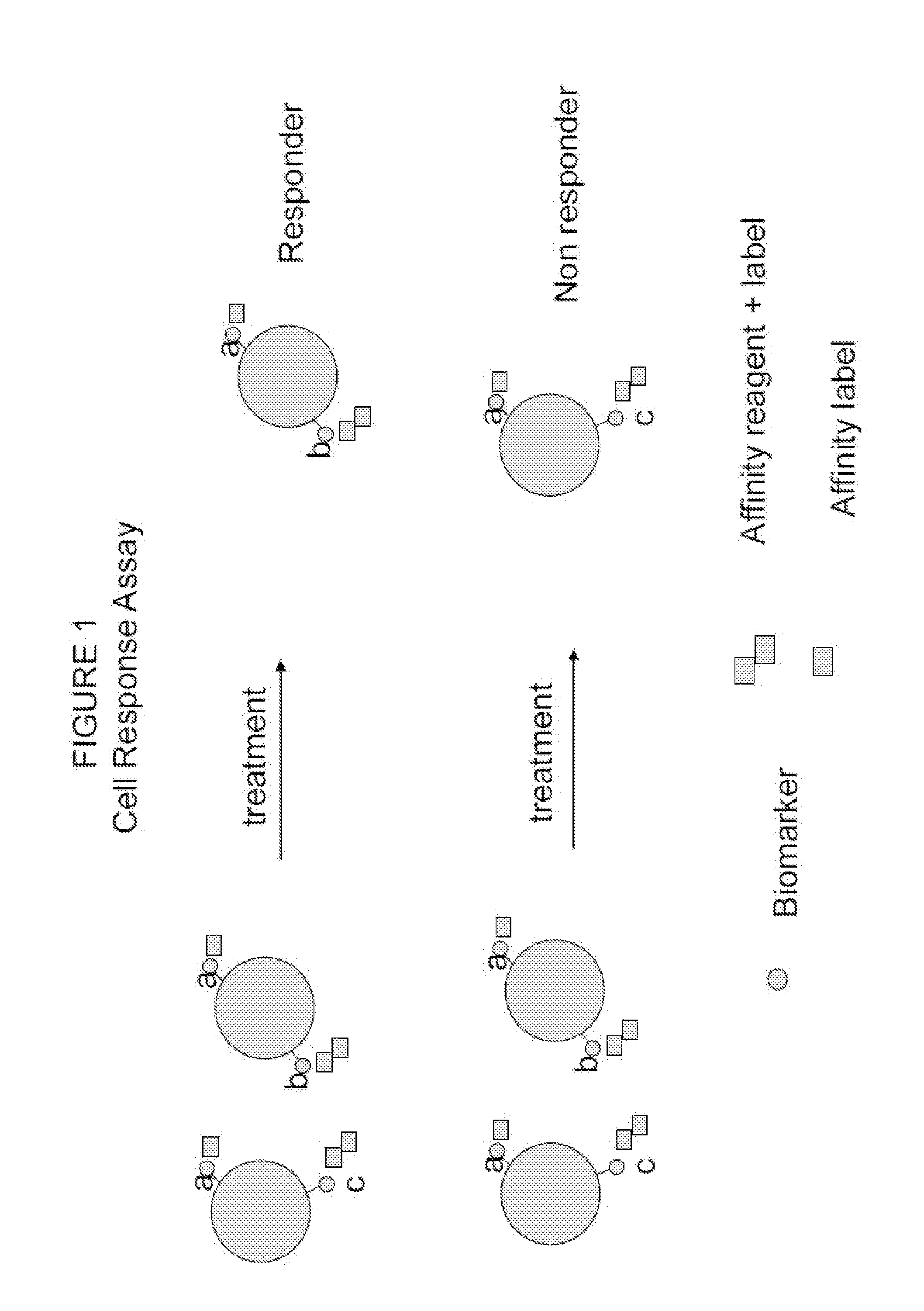
[0079]The following procedure shows the multiplexing of Biomarkers A (cancer cell type), B (metastatic potential) and C (chemo resistance) using a unique combination of affinity reagents with fluorescent labels and an affinity label to provide a multiplexed assay for breast cancer. This allows the simultaneous measurement of cancer cell type, chemo resistance nature, and metastatic potential, as described herein above, in combination with a marker indicating the presence of cell nucleus (utilizing a nucleic acid binding probe). The data demonstrated that four simultaneous signals can be measured with this combination but not with the traditional combinations of the prior art. The presently disclosed and claimed inventive concept(s) has the potential to measure up to eight signals utilizing various fluorophores taught herein.

[0080]The procedure to test the method was to collect 4 mL normal whole blood fresh into a VACUTAINER® tube (BD 6 mL VACUTAINE...

example 2

Cell Response Assay for Breast Cancer

[0085]A novel cell response assay for breast cancer was developed by using HER2 / neu as a tissue type marker for carcinoma breast cancer (Biomarker A) that is always present in the disease state. Biomarker B for metastatic potential utilized uPA and PAI antigen; these are markers of tumor invasiveness and are present in aggressive cancer cell differentiation and growth and increase with metastatic invasiveness potential of the cells. Biomarker C for chemo resistance utilized PL2L piwi like antigen (see for example, Gao et al., 2008).

[0086]The procedure shown in Example 1 was used to measure cancer cells before and after treatment with and without camptothecin. Camptothecin is known to activate apoptosis in cancer cells and kill cancer cells like a chemotherapy agent (See Gupta, 1997). Cells were tested by the cell response assay and by a traditional prior art assay. The novel “cell response assay” utilized HER2 / neu as Biomarker A for cell type, th...

example 3

Multiplexed Assay for Prostate Cancer

[0089]The following procedure shows the multiplexing of Biomarkers A (cancer cell type), B (metastatic potential) and C (chemo resistance) using a unique combination of affinity reagents with fluorescent labels and an affinity label to provide a multiplexed assay for prostate cancer. This allows the simultaneous measurement of cancer cell type, chemo resistance nature, and metastatic potential, as described herein above, in combination with a marker indicating the presence of cell nucleus (utilizing a nucleic acid binding probe). The data demonstrated that four simultaneous signals can be measured with this combination but not with the traditional combinations of the prior art. The presently disclosed and claimed inventive concept(s) has the potential to measure up to eight signals utilizing various fluorophores taught herein.

[0090]The procedure to test the method was to collect 4 mL normal whole blood fresh into a VACUTAINER® tube (BD 6 mL VACUT...


PUM

PropertyMeasurementUnit
Electrical resistanceaaaaaaaaaa
Wavelengthaaaaaaaaaa

Abstract

A cell response assay for cancer is provided. In the assay, the levels of a cancer cell type biomarker, a chemo resistance biomarker and a metastatic potential biomarker are simultaneously measured in a biological sample.

Description

CROSS REFERENCE TO RELATED APPLICATION[0001]The present application is based on provisional application Ser. No. 61 / 538,302, filed Sep. 23, 2011, the entire contents of which are herein incorporated by reference.STATEMENT REGARDING FEDERALLY SPONSORED RESEARCH OR DEVELOPMENT[0002]Not Applicable.BACKGROUND OF THE INVENTION[0003]1. Field of the Invention[0004]The presently disclosed and claimed inventive concept(s) generally relates to cellular assays. More particularly, but not by way of limitation, the presently disclosed and claimed inventive concept(s) relates to methods of assaying biological samples to determine the cellular response to treatment / therapy and / or monitor progression of a disease state.[0005]2. Description of the Background Art[0006]Affinity assays using fluorescence compounds are commonly used for cell and tissue analysis for biomarkers. The fluorescent compounds (labels) are attached (conjugated) to affinity molecules. Said affinity molecules may be, for example ...

Claims


Application Information

Patent Timeline
IPC IPC(8): G01N33/574
CPCG01N33/57484G01N33/57434G01N33/57415G01N33/5748G01N33/57407G01N2800/60C12Q1/6886C12Q2600/158C12Q2537/143C12Q2565/102A61K31/4745G01N2333/8132G01N2333/82G01N2333/91205G01N2333/96463
Inventor PUGIA, MICHAEL
Owner SIEMENS HEALTHCARE DIAGNOSTICS INC
Learn More