System and Device for Tumor Characterization Using Nonlinear Elastography Imaging

a nonlinear elastography and tumor technology, applied in the field of human tumor noninvasive classification, can solve the problems of low specificity rate, sensitivity decline, and imaging method's disadvantage of low sensitivity and specificity rate when used alon

Active Publication Date: 2014-09-25
WEST VIRGINIA UNIVERSITY
View PDF8 Cites 15 Cited by
  • Summary
  • Abstract
  • Description
  • Claims
  • Application Information

AI Technical Summary

Benefits of technology

[0037]Another embodiment of this invention provides a device, as described herein, including a force gauge that is attached to and in communication with the 3D ultrasound transducer probe for providing continuous force measurements, which enables a smooth force-strain measurement.

Problems solved by technology

1. X-ray imaging: This method uses ionizing radiation, which is unfavorable with pregnant patients. For breast masses, mammography is the current clinical imaging modality for breast masses. It has high sensitivity for detecting tumors, yet for patients with dense tissues, the sensitivity declines.
2. Conventional ultrasound imaging: this imaging method has the disadvantage of the low sensitivity and specificity rates when used alone.
3. Magnetic resonance imaging: has a good sensitivity rates for detecting malignancy. It has the disadvantages of lower specificity rates, being cost inefficient and not suitable with patients having metal implants.
4. Computed tomography: This method uses ionizing radiation, which is unfavorable with pregnant patients. It has good contrast with the ability to show tumor margins. However this method suffers from technological issues that need solving before applying it clinically.
5. The routine use of biopsy procedures to obtain tissue samples and histologically diagnose tumors. This method has the disadvantage of being invasive.
6. The use of strain imaging in the classification of tumors based on a linear elastic tissue model. These methods have the disadvantage of an existing overlap between the classification parameters used.
7. The use of shear wave elastography to visualize tumors and classify based on the estimated elasticity parameters. It has the disadvantage of not being able to diagnose very hard tumors.

Method used


Image

  • System and Device for Tumor Characterization Using Nonlinear Elastography Imaging
  • System and Device for Tumor Characterization Using Nonlinear Elastography Imaging
  • System and Device for Tumor Characterization Using Nonlinear Elastography Imaging

Examples


Embodiment Construction

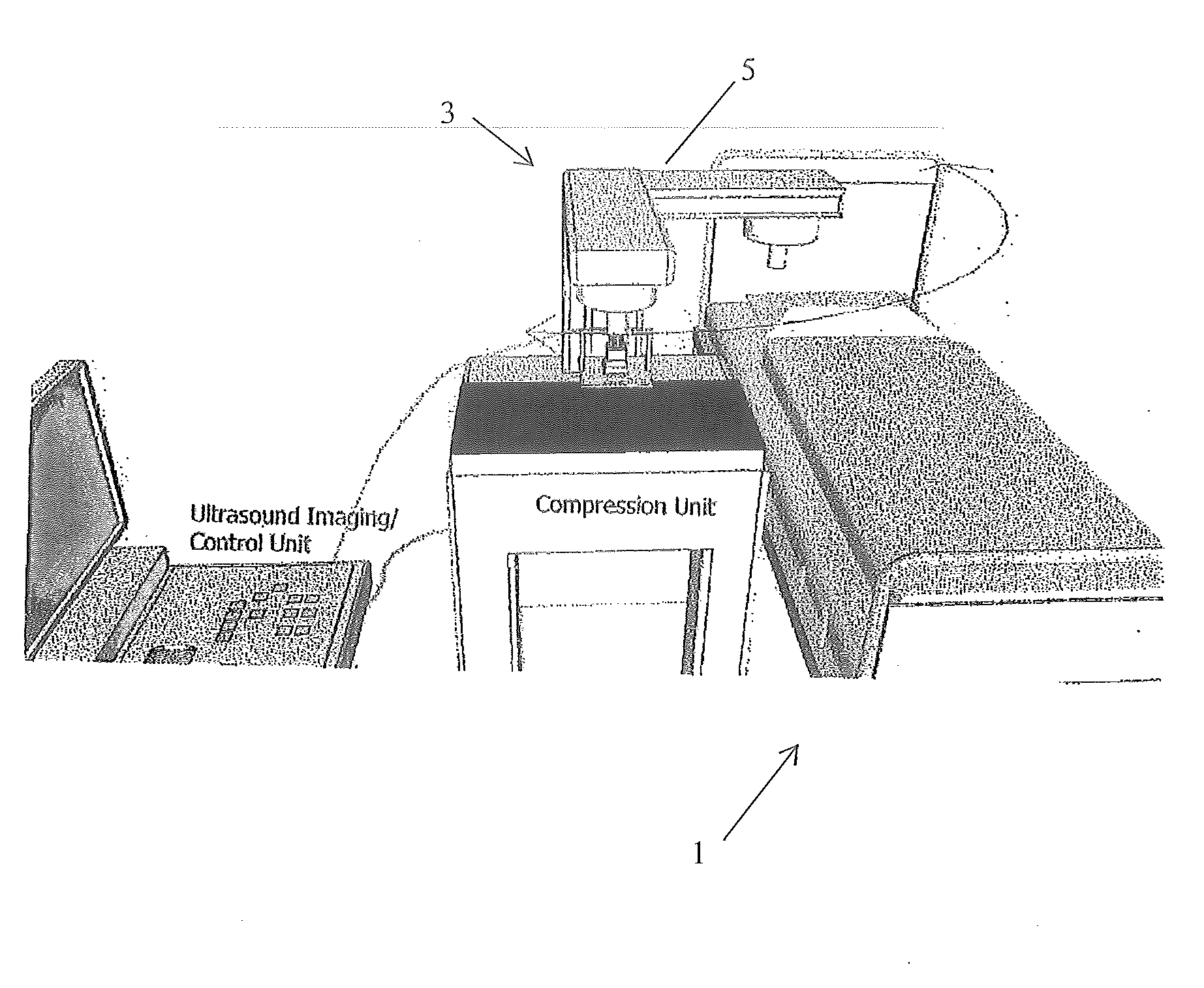
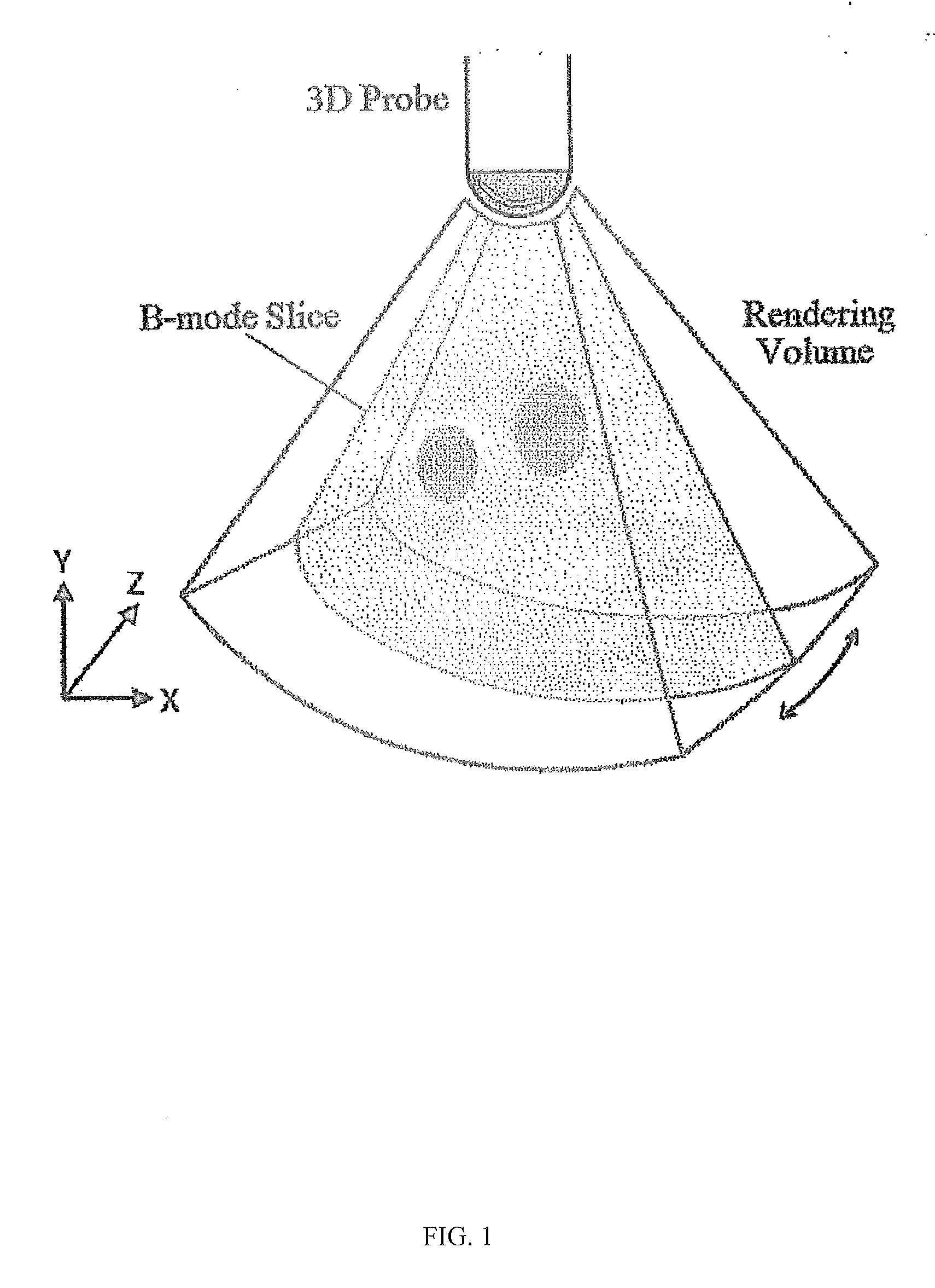
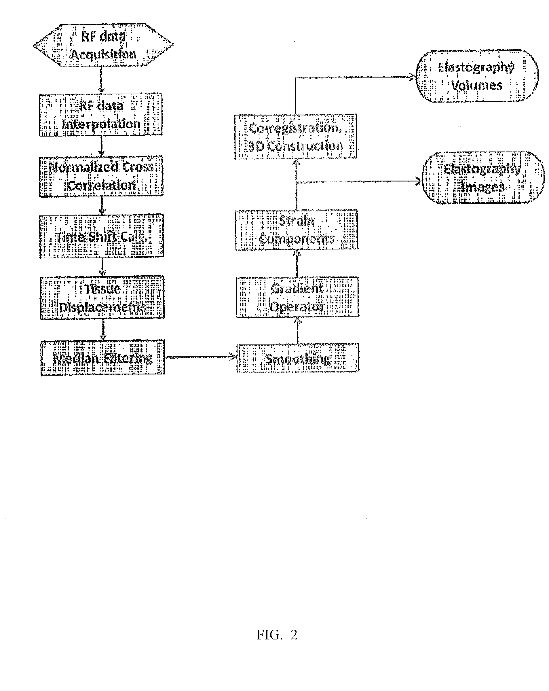
[0087]The present invention provides a method for classifying and characterizing a tumor of a patient as either benign or malignant comprising positioning a tissue or organ of a patient on a compression stage, aligning a 3D (three dimensional) ultrasound probe on or in the vicinity of the tissue or organ suspected of having a tumor, the probe capable of performing 3D ultrasound strain imaging (elastography), applying a first compression force to the tissue or organ having the suspected tumor for forming a first compressed tissue or organ, performing 3D ultrasound strain imaging (elastography) to the first compressed tissue or organ for estimating tissue strain, applying a second compression force to the first compressed tissue or organ, wherein the second compression force is greater than the first compressive force for forming a second compressed tissue or organ, performing 3D ultrasound strain imaging (elastography) to the second compressed tissue or organ for estimating tissue st...


PUM


Abstract

The present invention provides a method and a device to image and characterize human tumors, and to classify the tumors as either malignant or benign. The method includes using a multi-compression technique upon the tissue or organ combined with a 3D ultrasound strain imaging of the compressed tissue or organ for acquiring raw data and analyzing the raw data using a computer processing unit equipped with a nonlinear biomechanical tissue model for tumor classification. A device is provided having a compression stage for delivering multi-compression with continuous force measurements, and a 3D ultrasound transducer strain imaging probe, wherein the imaging probe and the compression stage are in communication with a computer processing unit.

Description

CROSS-REFERENCE TO RELATED APPLICATION[0001]This utility patent application claims the benefit of priority to pending U.S. Provisional Patent Application Ser. No. 61 / 775,555, filed on Mar. 9, 2013. The entire contents of U.S. Provisional patent Application Ser. No. 61 / 775,555 is incorporated by reference into this utility patent application as if fully written herein.STATEMENT REGARDING FEDERALLY SPONSORED RESEARCH OR DEVELOPMENT[0002]This invention was made with government support under contract number 5R21DE019561 by the National Institute of Health (NIH). The government has certain rights in the invention.BACKGROUND OF THE INVENTION[0003]1. Field of the Invention[0004]The present invention provides method for non-invasive classification of human tumors using ultrasound elastography via constructing quantitative tissue strain images and volumes. A device is provided comprising a 3D ultrasound transducer and a compression stage or paddle, each of which are in communication with a c...

Claims


Application Information

Patent Timeline
Patent Type & Authority Applications(United States)
IPC IPC(8): A61B8/00A61B8/08
CPCA61B8/403A61B8/485A61B8/4494A61B8/5207A61B8/483A61B8/085A61B8/4483A61B8/5223G16H50/30
Inventor MUKDADI, OSAMA M.ABDULHAMEED SAYED, AHMED M.LAYNE, GINGER P.ABRAHAM, JAME
Owner WEST VIRGINIA UNIVERSITY
Why Eureka
Try Eureka