
Capturing of cell fluid and analysis of its components under observation of cells and instruments for the cell fluid capturing and the analysis

a cell fluid and analysis technology, applied in the field of real-time capturing of cellular fluid and analysis of its components, can solve the problems of not being able to detect huge quantities of molecular groups and ions in living cells in real time or in time sequences, and achieve the effect of clarifying molecular mechanisms and confirming causes

Active Publication Date: 2014-10-23
HUMANIX
View PDF0 Cites 61 Cited by
  • Summary
  • Abstract
  • Description
  • Claims
  • Application Information

AI Technical Summary

Benefits of technology

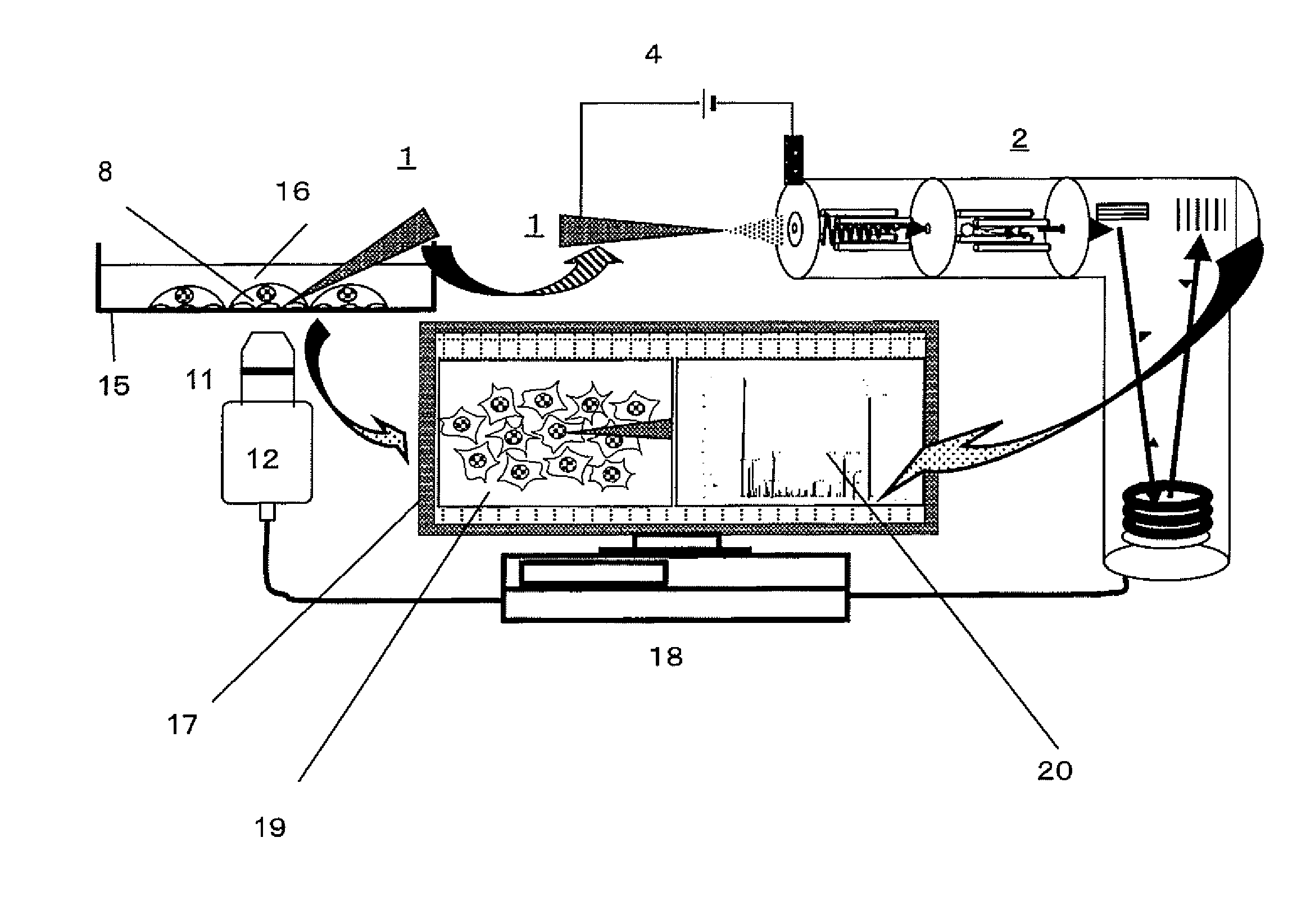
The invention provides a method to capture the contents of single cells and sub-cellular organelles in real-time, with high sensitivity and rapid detection. This allows for molecular analysis and the observation of cellular behaviors simultaneously. The method can be used in various fields such as medicine, food research, and chemical product industries to better understand the causes of disorders and improve quality assurances. The detection of ultralow amounts of substances in single cells or sub-cellular organelles can also be achieved.

Problems solved by technology

However, by present, there has been no analytical method to detect huge quantities of molecular groups and ions in living cells in real time or in time sequences with simultaneous direct observation of the cells, and then the methods identify and explore the key molecules to clarify the molecular mechanisms associated with observed phenomena, such as morphology.

Method used


Image

  • Capturing of cell fluid and analysis of its components under observation of cells and instruments for the cell fluid capturing and the analysis
  • Capturing of cell fluid and analysis of its components under observation of cells and instruments for the cell fluid capturing and the analysis
  • Capturing of cell fluid and analysis of its components under observation of cells and instruments for the cell fluid capturing and the analysis

Examples


Embodiment Construction

IN APPLICATION OF THIS INVENTION

[0047]The size of single cell ranges from 0.1 micrometer in diameter in the small one to enough visible large one such as eggs. Capturing of cell contents of nerve cells and eggs are easy which have large intracellular volumes and contain a lot of various components. Thus the principal target for solution of problems is the usual cell size about 10 micrometers in diameter from which it is difficult to capture the intracellular components. Then the analytical method can universal to almost all cases, if we care the case of 10 micrometers one.

[0048]However, the intracellular volume is less than 1 pico-liter and the volume of sub-cellular organelle is less thank 1 / 10 of it. Thus, the detection limit of current mass spectrometer is around 1 million numbers of molecules in each, contained in a single cell. It has been very difficult in targeting it from the aspect of current sensitivity. The manipulation in such micro space and ionizations, detections and ...


PUM


Abstract

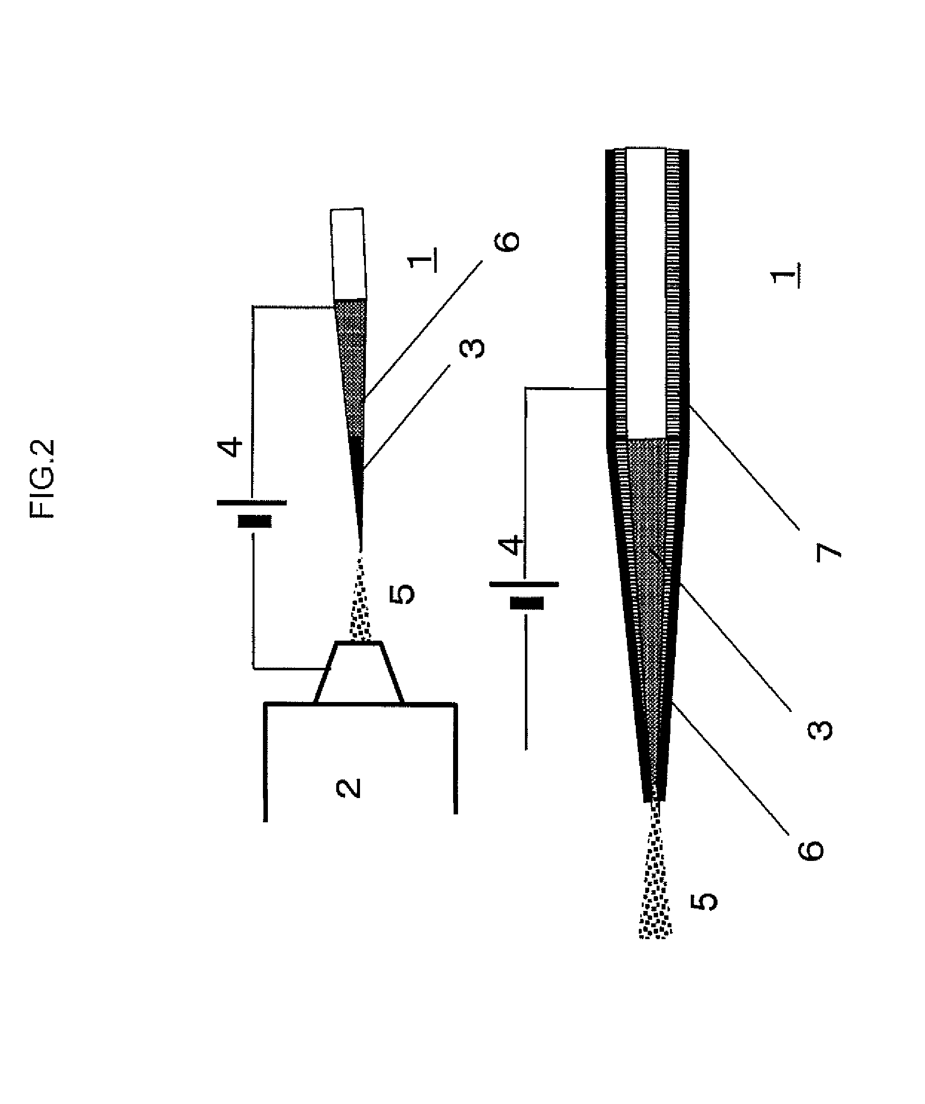
A method captures cellular components from a single cell and performs mass spectrometry on the components. The method includes inserting a nanospray ionization capillary tip into a specific region of the cell under observation with a microscope. The nanospray ionization capillary tip can include a filament in the interior. The method further includes capturing the cellular components of the specific region of the cell into the opening of the nanospray ionization capillary tip and keeping the components at the nanospray ionization capillary tip, supplying an ionization supporting solvent from a back-end of the nanospray ionization capillary tip, applying an electric field between a sample inlet of a mass spectrometer and the nanospray ionization capillary tip, whereby nanospray ionization to the cellular components is implemented, and performing the mass spectrometry on the cellular components captured at the nanospray ionization capillary tip.

Description

CROSS-REFERENCE TO RELATED APPLICATIONS[0001]This application is a continuation in part of U.S. application Ser. No. 12 / 739,660, filed on Aug. 30, 2010, as the U.S. National Phase under 35 U.S.C. §371 of International Application PCT / JP2008 / 070060, filed Nov. 4, 2008, which claims priority to Japanese Patent Application No. 2007-286208, filed Nov. 2, 2007, and Japanese Patent Application No. 2008-095092, filed Apr. 1, 2008. All of these related applications are hereby incorporated by reference in their entirety.FIELD OF THE INVENTION[0002]This invention is relates to real-time capturing of cellular fluid and analyses of its components under the cell observation and instruments for the cell-fluid capturing and the analyses.BACKGROUND OF THE INVENTION[0003]Molecular mechanisms of cells are the essential of life phenomena, and they are eternal subjects to be clarified in life sciences. However, by present, there has been no analytical method to detect huge quantities of molecular group...

Claims


Application Information

Patent Timeline
Patent Type & Authority Applications(United States)
IPC IPC(8): H01J49/04
CPCH01J49/0404Y10T436/24H01J49/167G01N27/62G01N33/48H01J49/04
Inventor MASUJIMA, TSUTOMUTSUYAMA, NAOHIROMIZUNO, HAJIME
Owner HUMANIX
Learn More