

Methods for preserving photoreceptor cell viability following retinal detachment
a technology of photoreceptor cells and viability, which is applied in the field of preservation of the viability of photoreceptor cells following retinal detachment, can solve the problems of partial or even complete blindness, deprived of blood and nutrients, sensitive photoreceptor cells disposed in the detached portion of the retina
- Summary
- Abstract
- Description
- Claims
- Application Information
AI Technical Summary
Benefits of technology
Problems solved by technology
Method used
Image

Examples
example 1
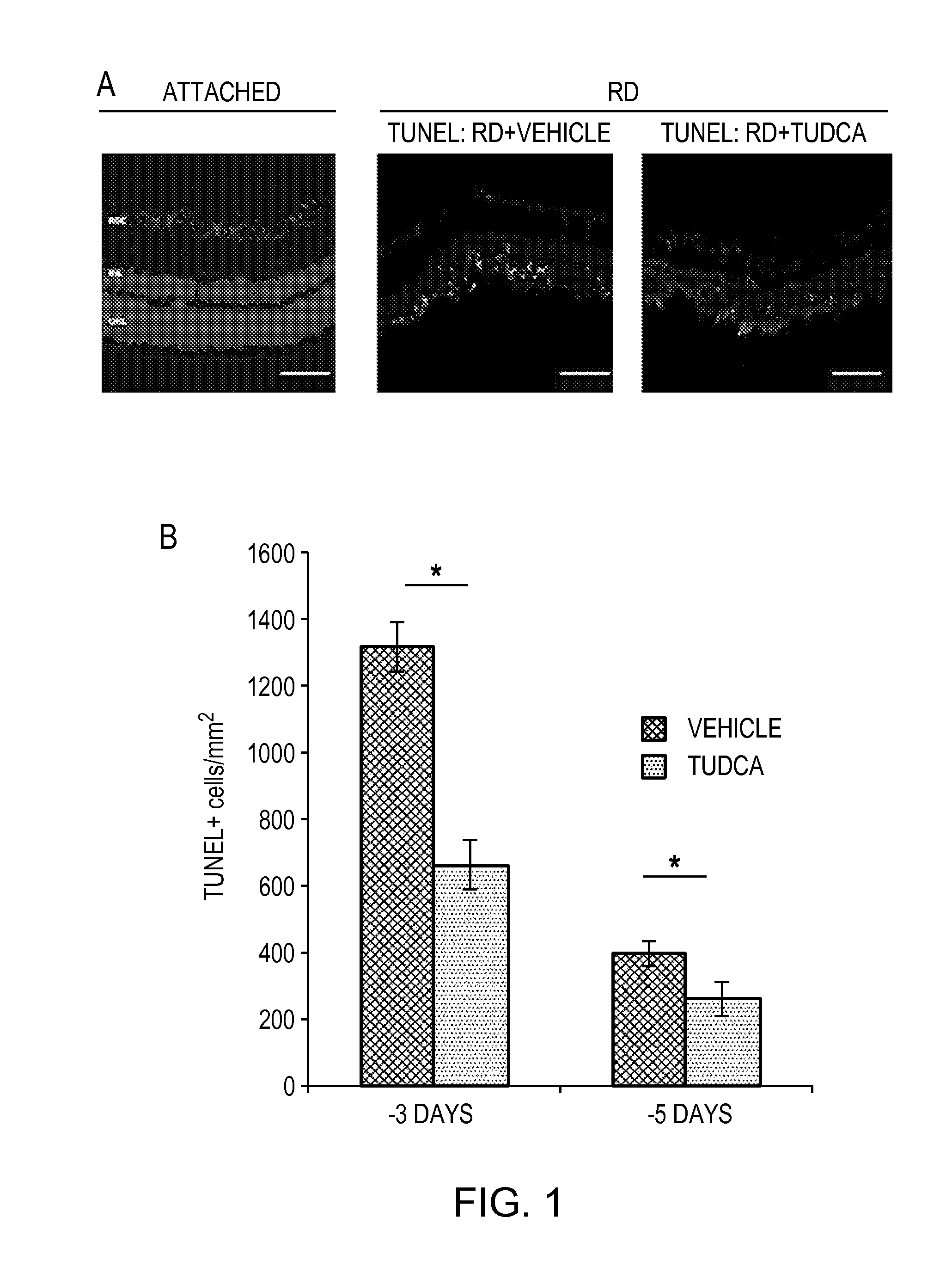
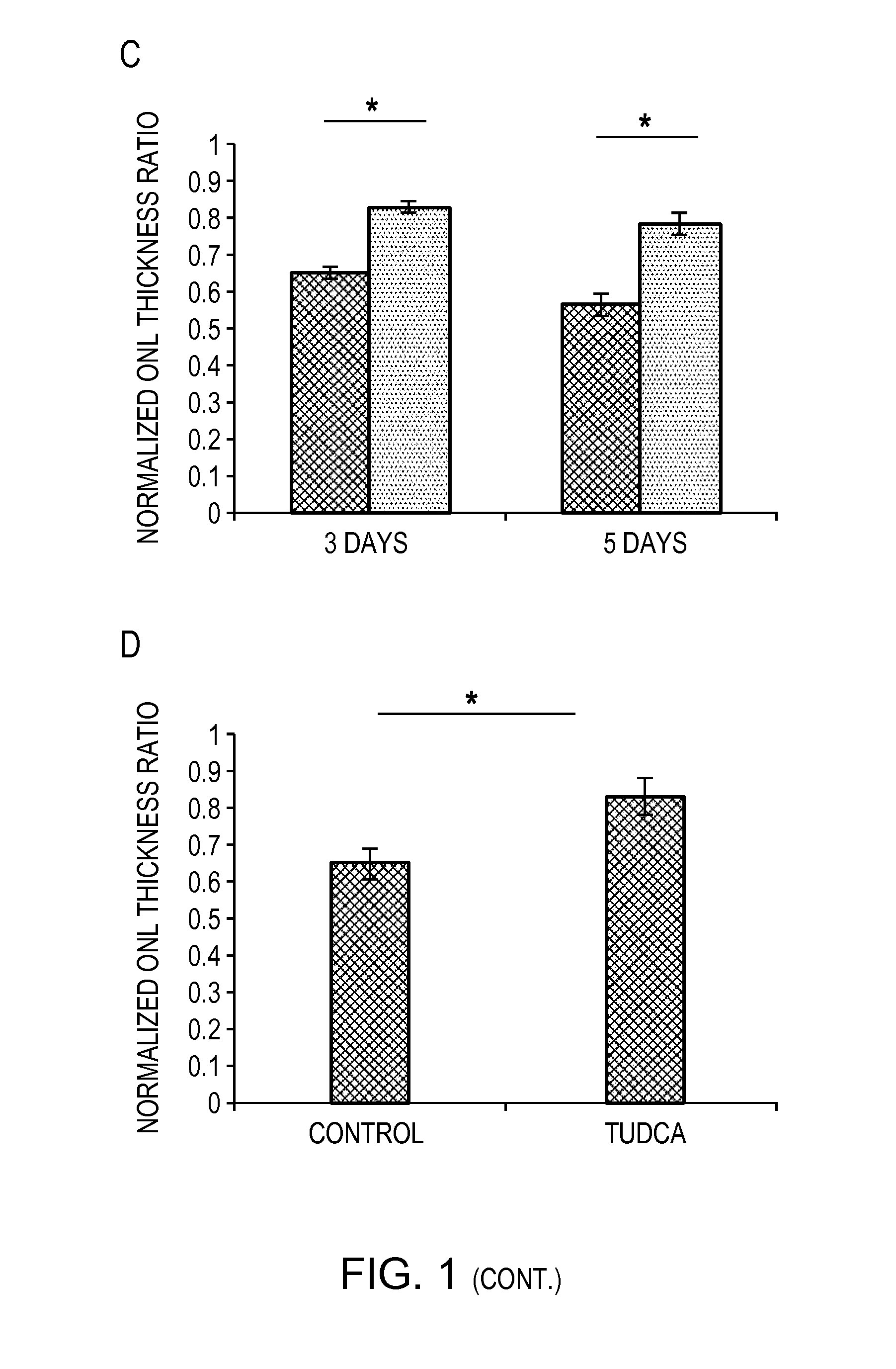
TUDCA Treatment Prevents Photoreceptor Death After Retinal Detachment
[0058]Photoreceptor death was assessed by TUNEL staining, which detects DNA fragmentation in apoptotic or necrotic nuclei. Rats were treated with either vehicle or TUDCA (500 mg / kg / day) injected intraperitoneally starting at 24 hours prior to the induction of retinal detachment. The rats were euthanized at three days or five days after induction of retinal detachment. The eyes were enucleated and fixed in 4% paraformaldehyde (PFA) in phosphate buffer saline (PBS) at 4° C. overnight. Subsequently, the eyes were embedded in Optimal Cutting Temperature media (OCT—Tissue Tek; Sakura Finetec, Torrance, Calif.), frozen at −21° C., and cut into 10 μm thick sections. The sections were fixed in 4% PFA for fifteen minutes and subjected to DAPI nuclear staining (AnaSpec / Eurogentec Group) and Terminal dUTP Nick-End Labeling (TUNEL) staining according to manufacturer's instructions (Apop Tag Fluorescein Apoptosis detection kit,...
example 2
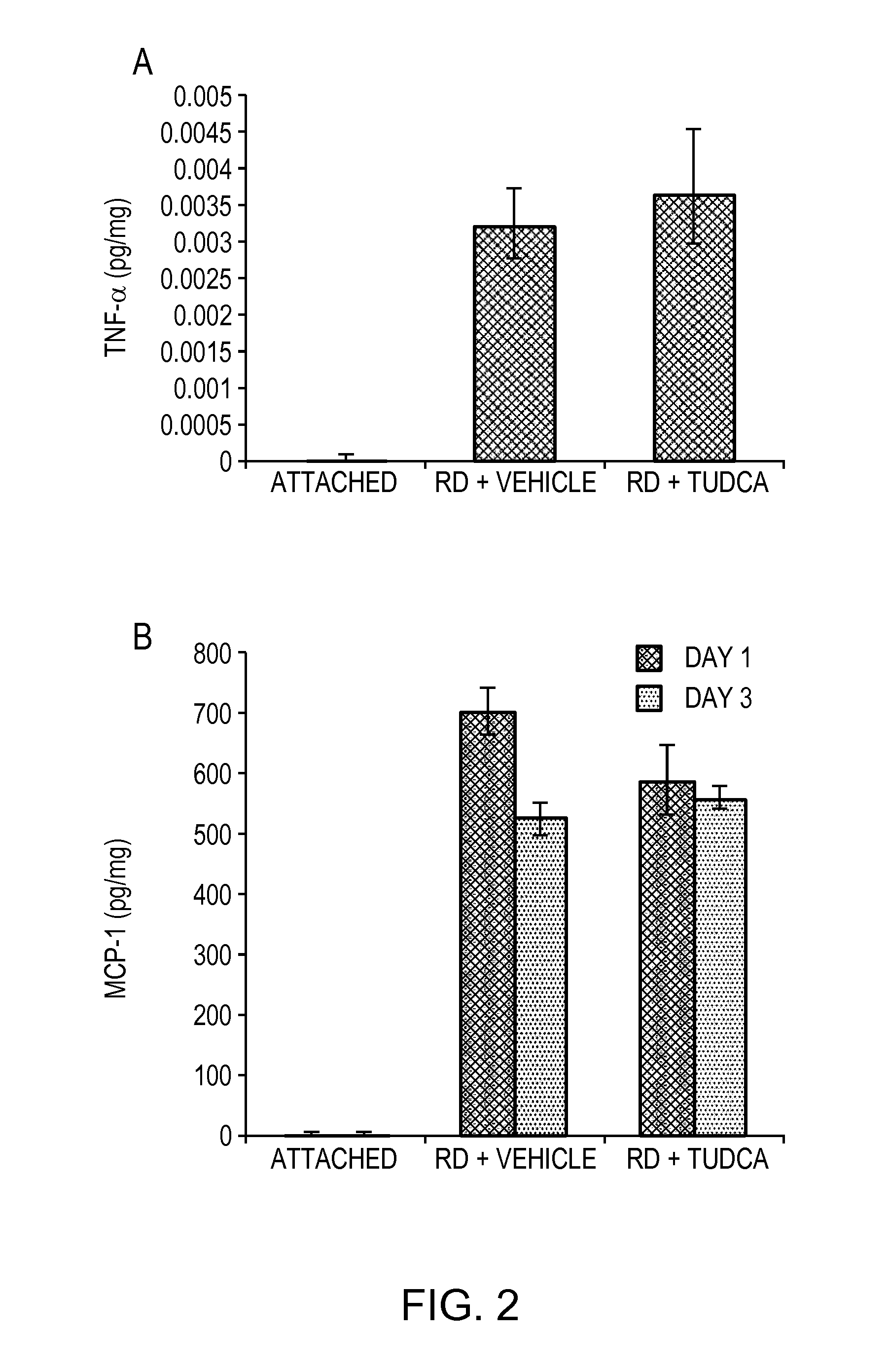
TUDCA Treatment Does Not Affect the Inflammatory Response Associated with Retinal Detachment
[0062]It has been shown previously that inflammatory cytokines and macrophage infiltration are markedly elevated in experimental retinal detachment (Nakazawa et al. (2007) PROC NATL ACAD SCI. 104: 2425-2430; Nakazawa et al. (2006) MOL VIS. 12: 867-878; Trichonas et al. (2010) PROC NATL ACAD SCI. 107(50):21695-21700). More specifically, several studies have shown upregulation of inflammatory cytokines such as TNF-α and MCP-1 and the infiltration of macrophages in the detached retina.
[0063]In order to assess macrophage infiltration in detached retinas, six eyes from each group (rats treated with vehicle or TUDCA) were analyzed. Eye sections were obtained as previously described, fixed in 4% PFA-PBS overnight, blocked in skim milk, and incubated overnight with anti-CD68 monoclonal antibody (Millipore, clone ED-1, #MAB1435). After washing with PBS, the sections were incubated with a secondary ant...
example 3
TUDCA Treatment Reduces Oxidative Stress
[0066]TUDCA has been shown to exert cytoprotective effects in different models by reducing oxidative stress (Oveson et al. (2011) J NEUROCHEM. 116: 144-153; Wei et al. (2008) HUM MOL GENET. 17: 469-477). It has also been shown previously that retinal detachment leads to increased ROS products such as increased levels of protein carbonyl content (Trichonas et al. (2010) PROC NATL ACAD SCI. 107(50):21695-21700; Roh et al. (2011) INVEST. OPHTHALMOL. VIS. SCI.).
[0067]In order to assess the effect of TUDCA treatment on oxidative stress, the protein carbonyl content in the retina was determined Eight eyes from each group (animals without retinal detachment as well as animals with retinal detachment that received vehicle or TUDCA) were analyzed using ELISA assays. Each retina was dissected and lysed in 200 μl of protein lysis buffer (Vavvas et al. (1997) J BIOL CHEM. 272: 13255-13261), sonicated for ten seconds, and centrifuged at 4° C. for five minu...
PUM
 Abstract
Description
Claims
Application Information