Speckle and noise reduction in ultrasound images

Inactive Publication Date: 2014-12-04
EAGLEYEMED
View PDF6 Cites 2 Cited by
  • Summary
  • Abstract
  • Description
  • Claims
  • Application Information

AI Technical Summary

Benefits of technology

[0010]A handheld ultrasound imaging system and method includes features to reduce speckle and time gain compression noise. In one embodiment the handheld ultrasound system includes beam forming electronics and digital waveform generators to generate th

Problems solved by technology

However, the noise also presents other disadvantages, particularly in a telemedicine application.
Although there are different types of noise in ultrasound images, generally speaking the near field may have graininess caused by speckle noise.
Ultrasound images are thus inherently noisy and exhibit two major types of noise, speckle noise, time gain compression (TGC), and quantization noise.
In an ultrasound system the transmitted signal is rapidly attenuated in biological tissues and suffers a very large attenuation in a round trip.
As a result the TGC process introduces amplification of noise in a poor signal environment, which is then compounded by quantization noise.
These noise sources can significantly affect the image quality needed for diagnosis and also the compressability of ultrasound streams for network transport.
In particular, conventional ultrasound images have a hi

Method used


Image

  • Speckle and noise reduction in ultrasound images
  • Speckle and noise reduction in ultrasound images
  • Speckle and noise reduction in ultrasound images

Examples


Embodiment Construction

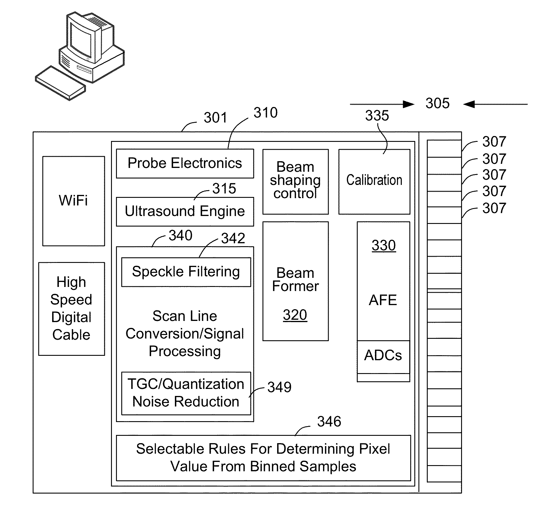
[0018]FIG. 3 is a block diagram illustrating aspects of an ultrasound imaging system in accordance with an embodiment of the present invention. The ultrasound imaging system may be used to transmit a live video stream of ultrasound images over a network for real-time review by another doctor. Thus, image quality and compressibility are important considerations.

[0019]In one embodiment the ultrasound imaging system is implemented as a hand held ultrasound system including electronics to generate the transmitted ultrasound pulses in a firing sequence and electronics to receive and process the reflected ultrasound pulses. In one embodiment the hand held ultrasound system includes a housing 301, a detachable transducer array 305 having an array of transducer elements 307, such as an array of piezoelectric crystals. The handheld ultrasound system may have a housing 301 that is probe shaped. It will also be understood that the handheld ultrasound system of the present invention may have a ...


PUM


Abstract

An ultrasound imaging system includes features to reduce speckle and time gain compression noise. A handheld ultrasound system may include beam forming electronics and digital waveform generators to generate the transmitted pulses with fine grained apodization to improve coherence and reduce speckle. Speckle filtering may be included in the ultrasound system. Features to reduce quantization noise and improve the time gain compression response may be provided.

Description

CROSS REFERENCE TO RELATED APPLICATIONS[0001]The present application claims the benefit of U.S. Provisional Application No. 61 / 829,891, filed on May 31, 2013, the contents of which are hereby incorporated by reference.FIELD OF THE INVENTION[0002]The present invention is generally related to techniques to reduce noise and improve image quality in ultrasound medical images.BACKGROUND OF THE INVENTION[0003]FIG. 1 illustrates an ultrasound medical image. Noise in ultrasound medical images presents several different aspects. Some types of noise can enhance the visual contrast between tissues. However, the noise also presents other disadvantages, particularly in a telemedicine application.[0004]Although there are different types of noise in ultrasound images, generally speaking the near field may have graininess caused by speckle noise. The far field is may have noise associated with time gain compression (TGC) and quantization noise.[0005]Ultrasound images are thus inherently noisy and e...

Claims


Application Information

Patent Timeline
IPC IPC(8): A61B8/08A61B8/14A61B8/00
CPCA61B8/5269A61B8/145A61B8/5207A61B8/4488B06B1/0215G01S7/52023G01S7/52046G01S7/52077G01S15/8915G10K11/346G01S7/5208A61B8/14
Inventor HIRIYANNAIAH, HARISH P.
Owner EAGLEYEMED
Why Eureka
Try Eureka