MRI Transfer Table Assembly

a transfer table and assembly technology, applied in the field of magnetic resonance imaging equipment, can solve the problems of not routinely performing magnetic resonance imaging (mri), infants must be moved, and the infant cannot be moved

Active Publication Date: 2014-12-11
CHILDRENS HOSPITAL MEDICAL CENT CINCINNATI
View PDF1 Cites 40 Cited by
  • Summary
  • Abstract
  • Description
  • Claims
  • Application Information

AI Technical Summary

Benefits of technology

[0011]A transfer table assembly is disclosed that is suitable for use in association with an MR scanner and an incubator for neonatal infants. The transfer table assembly generally includes a transfer table base that supports a transfer table. The transfer table may be removably connected with the top surface of the transfer table base. An associated incubator may include a bed for supporting a neonate when being cared for in the incubator. The transfer table may be configured to receive the incubator bed and the patient. In order to provide additional support and stability, the transfer table assembly may include an attachment mechanism for releasably attaching the transfer table base to the incubator in such a manner that said transfer table is substantially at the same level of the incubator bed. Additionally, the transport table assembly may include a supporting mechanism for providing additional structural support to the transfer table base when the transfer table base is attached to the incubator.

Problems solved by technology

Magnetic resonance imaging (MRI) is a powerful technique for diagnostic purposes but is not routinely performed on these infants due to a number of technical and logistical challenges.
One difficulty in utilizing MRI for these premature infants is monitoring the vital signs and life support of the infant during MR examination.
A second difficulty in utilizing MRI is that the infant must be moved from an incubator or isolette into and out of the MR scanner.
This movement places the infant at risk for injury.
At this stage, however, the infants are hemodynamically unstable.
Accordingly, transporting and maintaining homeostasis in these fragile infants presents difficulty.
Another challenge in using MRI for neonates is that MRI systems are frequently located in Radiology departments outside of and perhaps distant from the NICU.
This may present certain logistical and technical challenges with respect to controlling the neonate's environment.
Furthermore, removing staff from the NICU to transfer and attend to one baby outside the NICU can place the remaining babies in the NICU at increased risk of a reduced level of care due to decreased staff coverage.
While this approach has the advantage of not disturbing the baby while it is in the transport incubator, even during MR scanning, it has several limitations including: a) infants must be fully detached from the monitoring equipment in their home incubator to be transferred into the transport incubator, b) the MR system that is used for imaging must have a bore large enough to accommodate the portion of the transport incubator containing the baby (thereby requiring a large heavy magnet), c) the baby and its attending staff need to leave the NICU for scanning, and d) because the transport incubator must be fully MR compatible while providing full life support for the baby, the system is heavy and expensive.
While this approach has the benefit of providing a magnet that is more easily installed in the NICU, it does have several limitations including the need for MR compatible incubators to be used throughout the NICU, or the transfer of a neonate from a non-MR-compatible home incubator to an MR-compatible incubator.
This approach also limits the access to the attending staff as they prepare the infant for MR scanning.

Method used


Image

  • MRI Transfer Table Assembly
  • MRI Transfer Table Assembly
  • MRI Transfer Table Assembly

Examples


Embodiment Construction

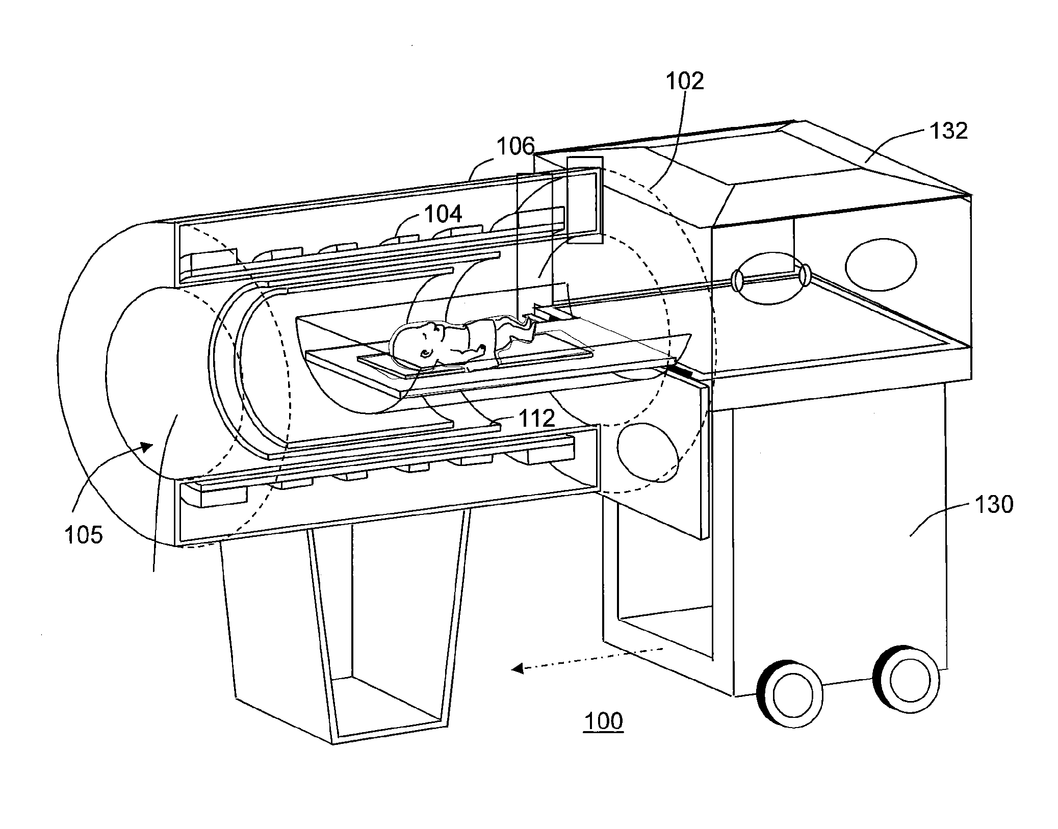
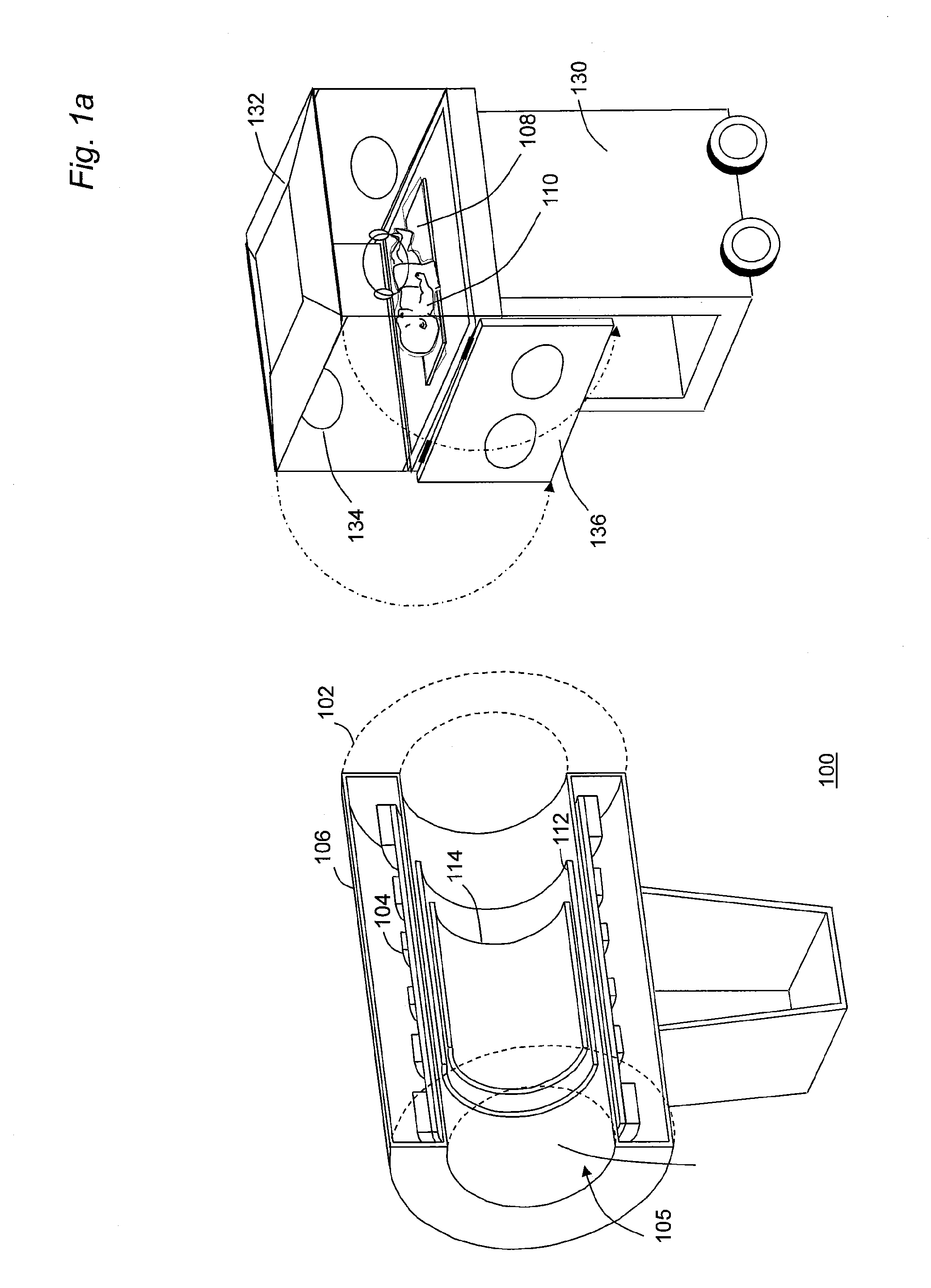
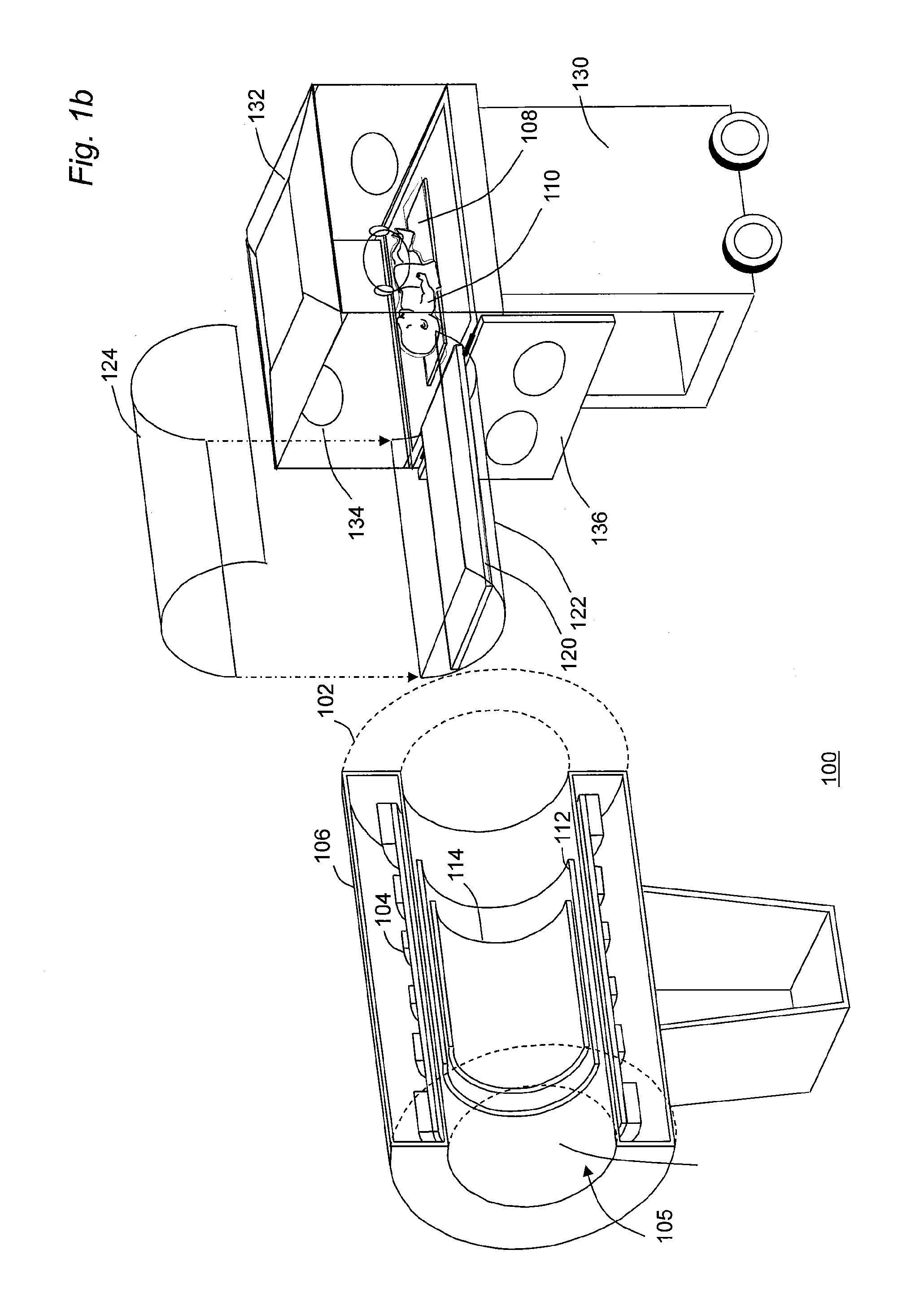
[0022]The use of MRI techniques for infants, and in particular neonates, is highly desirable. MRI techniques provide diagnostic information without patient exposure to ionizing radiation, and are suitable for extended and repeated studies.

[0023]MR techniques provide excellent anatomic visualization and functional information. They can be used to measure neural fiber track development and have a number of potential clinical uses including, but not limited, to diagnosis of brain trauma, cardiac abnormalities, congenital defects and the assessment of lung development.

[0024]There are, however, a number of challenges in the use of MRI for neonatal imaging. Patient access during scanning can be difficult as MR magnets are typically large and surround the patient. Safety concerns include forces on ferromagnetic objects, potential for rf heating and acoustic noise. Also, logistics may be difficult, as MR scanners tend to be in radiology departments, while neonate infants are typically in th...


PUM


Abstract

The present disclosure relates to a transfer table assembly suitable for use in association with an MR scanner. The transfer table assembly includes a transfer table attached to a transfer table base that may releasably engage an incubator. The transfer table may be dimensionally constructed such that it may be inserted directly into an MR scanner.

Description

CROSS-REFERENCE TO RELATED APPLICATIONS[0001]The present disclosure claims priority benefit of U.S. Provisional Patent Application Ser. No. 61 / 593,930 filed Feb. 2, 2012 and is further related to U.S. Provisional Patent Application Ser. No. 61 / 429,612 also filed Jan. 4, 2011.FIELD OF THE DISCLOSURE[0002]The present disclosure relates generally to magnetic resonance imaging equipment suitable for use in neonatal care and, more particularly, to a transfer table assembly for transferring a neonate from an incubator into an MR scanner.BACKGROUND OF THE DISCLOSURE[0003]Today premature infants weighing less than 1 kg may be stabilized and allowed to develop in neonatal intensive care units (NICUs). Magnetic resonance imaging (MRI) is a powerful technique for diagnostic purposes but is not routinely performed on these infants due to a number of technical and logistical challenges.[0004]When a mother has a high-risk pregnancy, it is possible that the baby could be born prematurely and would...

Claims


Application Information

Patent Timeline
Patent Type & Authority Applications(United States)
IPC IPC(8): A61B5/055G01G19/44A61B5/00A61G11/00
CPCA61B5/0555A61B5/0046G01G19/44A61G11/005A61B5/055A61B2503/045
Inventor DUMOULIN, CHARLES L.
Owner CHILDRENS HOSPITAL MEDICAL CENT CINCINNATI
Why Eureka
Try Eureka