Implantable Medical Devices, Methods of Use, and Apparatus for Extraction Thereof

a technology for implanted objects and medical devices, applied in the field of devices and methods for the removal of implanted objects, can solve the problems of increasing electrode resistance, fibrous scar tissue often forms, and the endocardial lead fails occasionally, so as to promote the release of the exterior surface

Inactive Publication Date: 2015-03-19
CHATURVEDI NEHA
View PDF0 Cites 23 Cited by
  • Summary
  • Abstract
  • Description
  • Claims
  • Application Information

AI Technical Summary

Benefits of technology

[0008]Embodiments of the present invention are directed to an implantable device, an extraction device and a method of using such implantable device and extraction device to extract the implantable device with a minimum of trauma to the animal or human in which the implantable device is placed. One embodiment directed to the implantable device for implantation into tissues of an animal or human comprises at least one body having an exterior surface for placement in intimate contact with a non-fluid tissue in which the device is implanted. The device further comprises at least one of the photoreactive agent, selected from the group consisting of a photo sensitizer or photosensitive nanoparticle, associated with the exterior surface. The delivery of light energy to the photoreactive agent generates at least one of the following forms of energy consisting of reactive compounds or thermal energy and promotes release of the exterior surface from the tissues in which it is implanted.

Problems solved by technology

In addition, fibrous scar tissue often forms, in part, over the insulator sleeve within the venous system and the heart chamber.
Although the state of the art in implantable pulse generator or pacemaker technology and endocardial technology has advanced considerably, endocardial leads nevertheless occasionally fail, due to a variety of reasons, including insulation breaks, breakage of the inner helical coil conductor thereof and an increase in electrode resistance.
Unused transvenous leads increase the risk complications will develop.
Possible complications associated with leaving unused leads in the heart and venous system include an increased likelihood an old lead may be the site of infection.
Development of an infection may, in turn, lead to septicemia, a possibly fatal complication.
Unused leads may also cause endocarditis.
Furthermore, unused leads may entangle over time, thereby increasing the likelihood of blood clot formation.
Such clots may embolize to the lung and produce severe complications or even fatality.
The presence of unused leads in the venous pathway and inside the heart can also cause considerable difficulty in the positioning and attachment of new endocardial leads in the heart.
Moreover, multiple leads within a vein or artery may impede blood flow causing fatigue, weakness or dizziness within the patient.

Method used


Image

  • Implantable Medical Devices, Methods of Use, and Apparatus for Extraction Thereof
  • Implantable Medical Devices, Methods of Use, and Apparatus for Extraction Thereof
  • Implantable Medical Devices, Methods of Use, and Apparatus for Extraction Thereof

Examples


Embodiment Construction

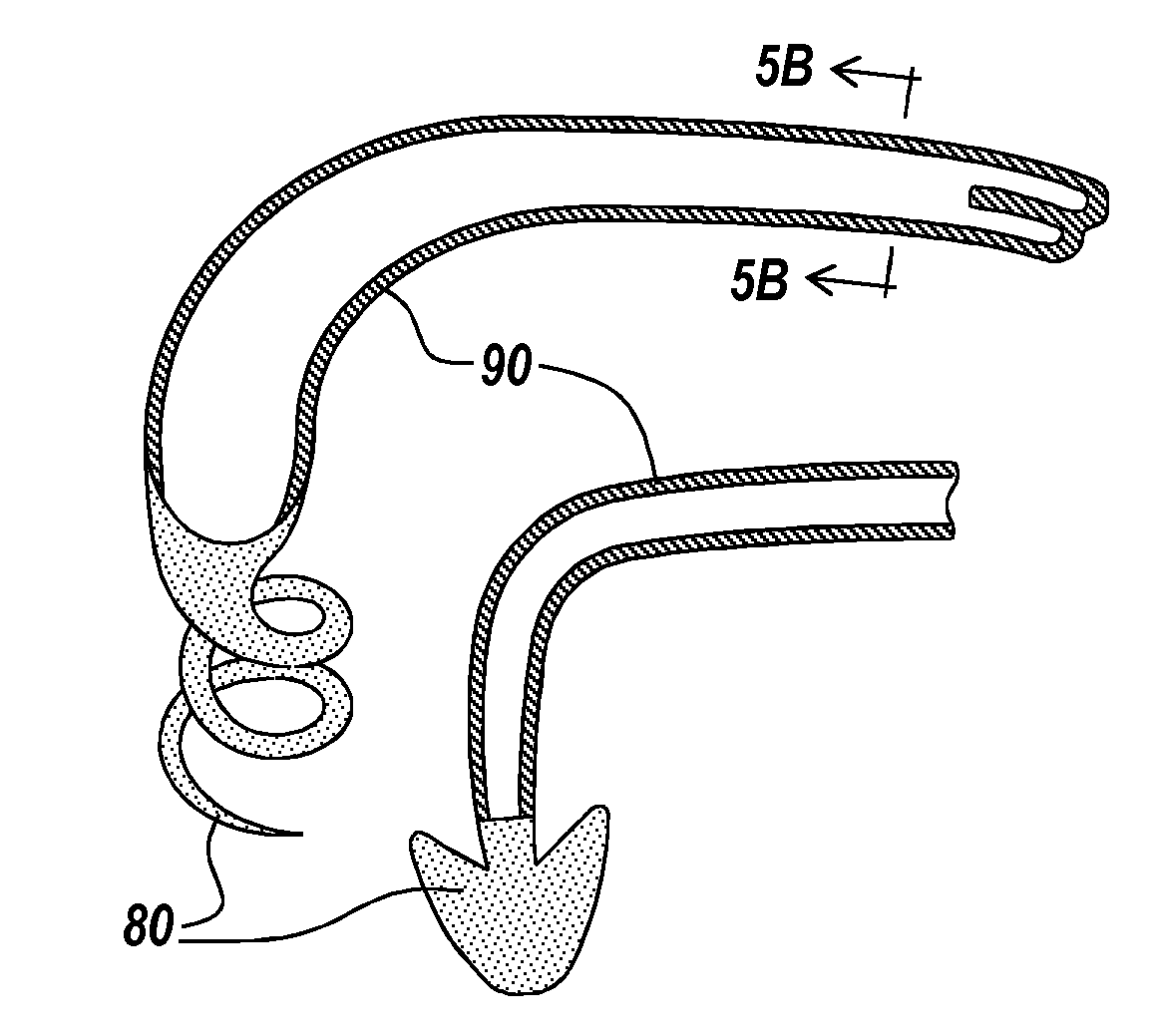
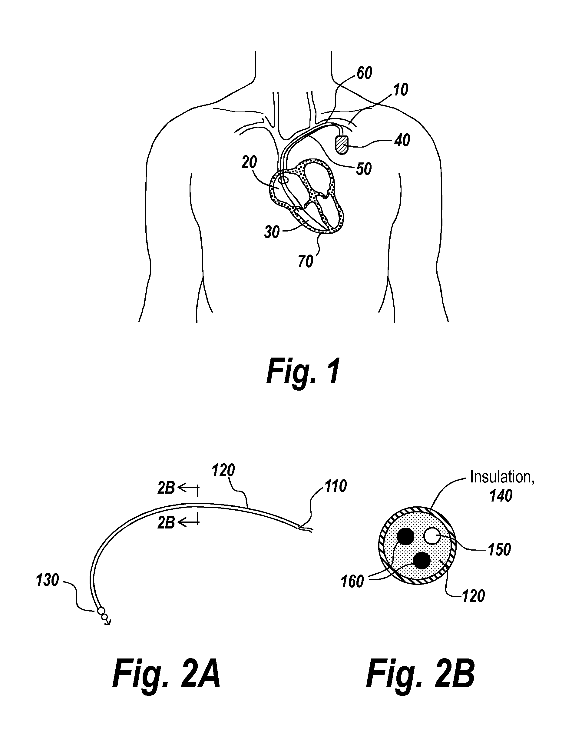
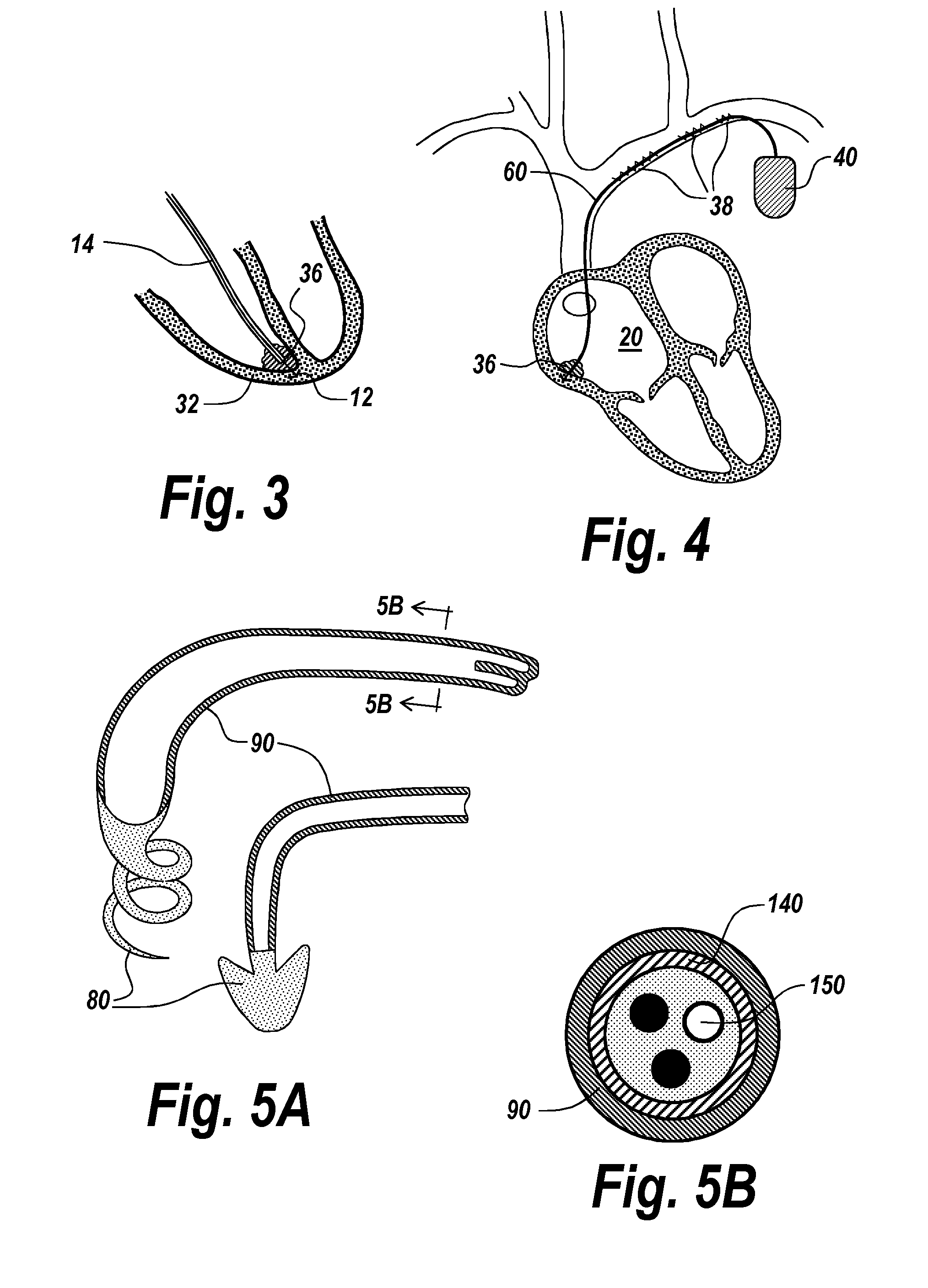
[0052]The present disclosure relates generally to devices and methods for removal of an implanted object from a subject's body, and more particularly to devices and methods for removal of endocardial leads from a patient's body using selected application of energy to the leads.

[0053]One aspect of the present disclosure relates an implantable medical device. The implantable medical device can include a main body portion having at least one photosensitive nanoparticle associated therewith. Delivery of energy to the main body portion promotes extraction of said implantable medical device from a subject.

[0054]Another aspect of the present disclosure relates to a method for extracting an implanted medical device from a subject. One step of the method includes identifying a medical device implanted in the subject. The medical device can comprise a main body portion including at least one photosensitive nanoparticle and / or photosensitizer associated therewith. Next, an amount of energy is ...


PUM


Abstract

One aspect of the present disclosure relates to an implantable medical device. The implantable medical device can include a main body portion having at least one photosensitive nanoparticle associated therewith. Delivery of energy to the main body portion promotes extraction of said implantable medical device from a subject.

Description

CROSS REFERENCE TO RELATED APPLICATIONS[0001]This application claims priority to U.S. Provisional Application Ser. No. 61 / 840,138 filed Jun. 27, 2013, the entire contents of which is incorporated herein by reference.STATEMENT REGARDING FEDERAL FUNDING[0002]Embodiments of the present invention were not conceived or developed with Federal funding or sponsorship.TECHNICAL FIELD[0003]The present disclosure relates generally to devices and methods for removal of an implanted object from a subject's body, and more particularly to devices and methods for removal of endocardial leads from a patient's body using selected application of energy to the leads.BACKGROUND[0004]Endocardial leads are often placed in contact with the endocardial tissue by passage through a venous access, such as the subclavian vein or one of its tributaries. Thus a transvenous endocardial lead refers to a pacemaker lead which contacts endocardial tissue through a vein. In the past, various types of transvenous endoca...

Claims


Application Information

Patent Timeline
Patent Type & Authority Applications(United States)
IPC IPC(8): A61B5/06A61B17/3205A61N1/05
CPCA61B5/065A61B17/3205A61N1/05A61N5/0601A61N5/062A61B18/24A61B2017/22087A61B2018/00351A61N2001/0578
Inventor CHATURVEDI, NEHA
Owner CHATURVEDI NEHA
Why Eureka
Try Eureka