Optical coherent imaging medical device

Active Publication Date: 2015-03-19
STRYKER EUROPEAN OPERATIONS LIMITED
View PDF26 Cites 17 Cited by
  • Summary
  • Abstract
  • Description
  • Claims
  • Application Information

AI Technical Summary

Benefits of technology

[0034]Levels of Absolute Perfusion vary greatly at different regions of the body and among individuals even when carefully stratifying patient groups by age, sex, BMI and the presence of microcirculatory disorder

Problems solved by technology

It is therefore difficult to set thresholds for Absolute Perfusion for specific assessments such as burns

Method used


Image

  • Optical coherent imaging medical device
  • Optical coherent imaging medical device
  • Optical coherent imaging medical device

Examples


example 1

Burns Healing Assessment

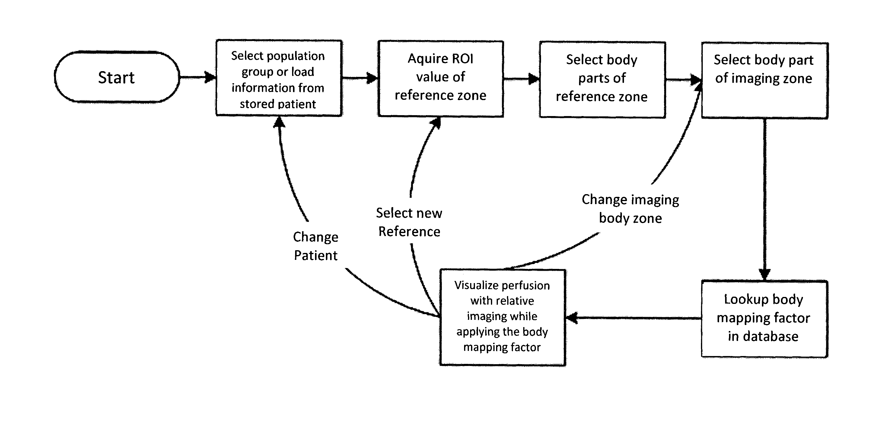
[0118]A workflow example is given for using Body Mapping Correction in the LDI based assessment of the healing potential of partial thickness burn wounds (see also FIG. 10). The principle here is to understand which parts of the burn wound are subject to an inflammatory response and thus hyperemic compared to equivalent skin at the same body area.[0119]1. Ask the user for the Population Group and then select the relevant Body Mapping Factor Table based on the Population Group (i.e. by taking age, skin pigmentation and comorbidities into account) and the Clinical Context (i.e. burn depth assessment). The information may be stored in the patient file.[0120]2. Take a Reference OCI Value on healthy skin. The device asks the user to select the body area where the reference is taken.[0121]3. The user is asked to select the body area where burn wound to be evaluated is located (Imaging Zone). If it is the same body area where the reference has been taken, then Body ...

example 2

Peripheral Vascular Disease (PVD) Assessment

[0125]A workflow example to identify and evaluate extremity perfusion impairment and post-interventional hyperemic response by comparing the forehead to extremity perfusion ratio with an appropriate Population Group without PVD is provided:[0126]1. Ask the user to select the Population Group, and then select the relevant Body Mapping Factor Table based on the Population Group (i.e. by taking comorbidities into account) and the Clinical Context (i.e. PVD).[0127]2. Take a Reference OCI Value (e.g. with LDI) on healthy skin (i.e. the forehead). The device asks the user to select or automatically sets the body area where the reference is taken.[0128]3. The user is asked to select the body area of the extremity to be evaluated, and the Body Mapping Factor is looked-up in the Body Mapping Factor Database to set the expected baseline perfusion level for the given Population Group.[0129]4. The (Relative) Perfusion Values and (Relative) Perfusion M...


PUM


Abstract

An OCI medical device includes a coherent light source, a light sensor, a first processing unit adapted to calculate OCI Data from the light sensor, a control unit which allows taking or loading of at least one Reference OCI Value, a second processing unit adapted to calculate the Intra-Individual Relative Assessment of the OCI Data of an Imaging Zone and the at least one OCI Reference Value, and display means adapted to show at least one Relative OCI Value. Uses and a method for assessing the blood flow of a body region use OCI imaging and include an Intra-Individual Relative Assessment between OCI Data of the Imaging Zone and at least one Reference OCI Value.

Description

FIELD OF INVENTION[0001]The invention relates to perfusion visualization using Optical Coherent Imaging (OCI) in the medical field.DEFINITIONS[0002]Optical Coherent Imaging (OCI): A contactless and non-invasive medical imaging modality utilizing the physical properties, and in particular the coherence properties, of light to record, measure, visualize and / or assess physiological and non-physiological flow properties, and in particular blood flow properties, in a selected tissue and / or body area of interest. OCI systems comprise of at least, but are not limited to: a coherent light source, a light acquisition unit, an image and data processing and device control unit, and means to visualize and represent the flow information. Specific implementations of OCI include, but are not limited to: Laser Doppler Imaging (LDI), Laser Doppler Spectroscopic Imaging (LDSI), Laser Speckle Imaging (LSI), Laser Speckle Contrast Analysis (LASCA), Functional Optical Coherent Imaging (fOCI, as for inst...

Claims


Application Information

Patent Timeline
IPC IPC(8): A61B5/026A61B5/00G06T7/00
CPCA61B5/0261G06T7/0014A61B5/0066G06T2207/30104A61B5/742G06T2207/10101A61B5/445A61B5/0059A61B5/0064A61B5/02007
Inventor ANDRE, MARCFRIEDRICH, MICHAELTHACHER, TYLERNAUMENKO, ANDREYFARKAS, ROMAIN
Owner STRYKER EUROPEAN OPERATIONS LIMITED
Why Eureka
Try Eureka