Intra-Operative Cancer Diagnosis Based on a Hyperpolarized Marker

a marker and intraoperative technology, applied in the field of intraoperative cancer diagnosis based on hyperpolarized markers, can solve the problems of invasion and metastasis, low likelihood of cancer spreading, and low specificity of the above-described preliminary histological investigation,

Inactive Publication Date: 2015-05-14
ALBEDA INNOVATION APS
View PDF1 Cites 1 Cited by
  • Summary
  • Abstract
  • Description
  • Claims
  • Application Information

AI Technical Summary

Benefits of technology

The present invention provides a method for diagnosing cancer in a tissue sample based on a hyperpolarized marker and NMR detection. This method can be carried out during surgery on a patient and can provide a reliable diagnosis of cancer in an excised tissue sample. The method involves contacting the tissue sample with at least one hyperpolarized marker, obtaining an NMR spectrum and / or MR image of the tissue sample, and comparing it to a reference. The method can be performed in a non-invasive manner and can be used in a specific patient population, such as patients with breast cancer. The hyperpolarized markers used can be fatty acids, amino acids, keto acids, TCA cycle intermediates, urea cycle intermediates, N-acetyl derivatives of amino acids, carbohydrates, 2-amino-phosphono-carboxylic acids, fluorinated alpha amino acids, quaternary nitrogen containing compounds, salts thereof, esters thereof, and mixtures thereof.

Problems solved by technology

Secondly, there is the problem of invasion and metastasis.
On the contrary, if no cancer cells are found in the sentinel lymph node, the likelihood of a spread of the cancer is low.
However, the specificity of the above-described preliminary histological investigation (such as H&E stained frozen sections, immunohistochemistry on frozen sections, imprint cytology and molecular analysis) is rather low.
The detection of metastases in this setup is limited to macrometastases due to a limited sensitivity.
All of the above methods have drawbacks, either in that they are only analyzing a minor fraction of the lymph node (e.g. about 1% for the H&E staining of frozen sections), or in that the assays correspond to destructive methods with regard to the analyzed tissue and do not allow for a reuse of the tissue for further types of diagnoses, such as e.g. a post-operative histopathology.
Summarizing the above, no reliable and non-invasive method of cancer diagnosis, which can be carried out during a surgical session, has been established to date.

Method used


Image

  • Intra-Operative Cancer Diagnosis Based on a Hyperpolarized Marker
  • Intra-Operative Cancer Diagnosis Based on a Hyperpolarized Marker
  • Intra-Operative Cancer Diagnosis Based on a Hyperpolarized Marker

Examples


example 1

3.1. Example 1

Detecting Cancer Metastases in Sentinel Lymph Nodes Through Differentiated Uptake of 15N Choline

[0243]Step 1: a sample of 15N choline, 0.05 mmol, is hyperpolarized according to procedures known in the art (Allouche-Arnon et al. Contrast Media Mol Imaging. 2011 November-December; 6(6):499-506).

[0244]Step 2: a sentinel lymph node from a prostate cancer patient undergoing prostate cancer surgery for a primary tumor in the prostate is identified, excised and rapidly (preferably within one minute from completed excision) placed in a 4 ml vessel filled with Ringer's solution tempered to 37° C.

[0245]Step 3: the vessel containing the lymph node is placed in an MR scanner which has been tuned to 15N and shimmed on a phantom resembling the vessel with the lymph node.

[0246]Step 4: the hyperpolarized marker is dissolved in 5 ml phosphate buffer (40 mM, pH 7.3) and otherwise prepared for use according to procedures known in the art (Ardenkjaer-Larsen et al. Proc Natl Acad Sci USA. ...

example 2

3.2. Example 2

Detecting Cancer Metastases in Sentinel Lymph Nodes Through Differentiated Uptake of 1-13C Acetate

[0251]Step 1: a sample of 1-13C acetate, 0.05 mmol, is hyperpolarized according to procedures known in the art (Jensen et al. J Biol Chem. 2009 Dec. 25; 284(52):36077-82).

[0252]Step 2: a sentinel lymph node from a breast cancer patient undergoing breast cancer surgery for a primary tumor in the breast is identified, excised and rapidly (preferably within one minute from completed excision) placed in a 2 ml vessel filled with Ringer's solution tempered to 37° C.

[0253]Step 3: the vessel containing the lymph node is placed in an MR scanner which has been tuned to 13C and shimmed on a phantom resembling the vessel with the lymph node.

[0254]Step 4: the hyperpolarized marker is dissolved in 5 ml phosphate buffer (40 mM, pH 7.3) and otherwise prepared for use according to procedures known in the art (Ardenkjaer-Larsen et al. Proc Natl Acad Sci USA. 2003 Sep. 2; 100(18):10158-63)....

example 3

3.3. Example 3

Detecting Cancer Metastases in Sentinel Lymph Nodes Through pH Differences

[0259]Step 1: a sample of 13C bicarbonate, 0.1 mmol, is hyperpolarized according to procedures known in the art (Gallagher et al. Nature. 2008 Jun. 12; 453(7197):940-3).

[0260]Step 2: a sentinel lymph node from a colon cancer patient undergoing colon cancer surgery for a primary tumor in the colon is identified, excised and rapidly (preferably within one minute from completed excision) placed in a 1 ml vessel filled with Ringer's solution tempered to 37° C.

[0261]Steps 3 to 4 are performed as in example 2.

[0262]Step 5: a fraction of the solution, 25 μl, containing the hyperpolarized marker is injected into the lymph node.

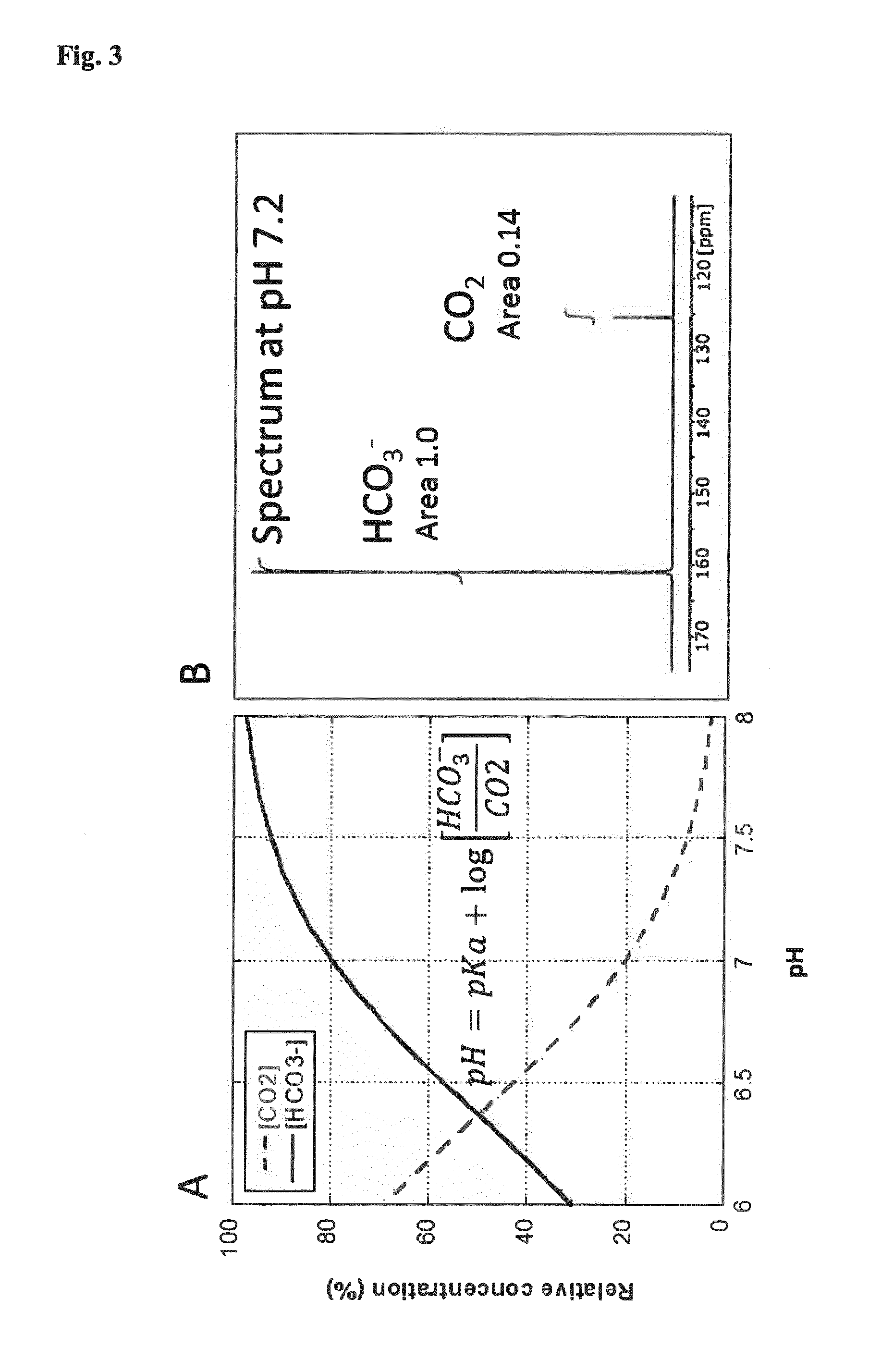
[0263]Step 6: after 10 to 30 s, a 13C MR spectroscopic investigation is performed on the lymph node and the signals from 13C bicarbonate and 13C carbon dioxide are quantified. The pH is calculated according to the Henderson-Hasselbalch equation (see FIG. 3) and compared to a standa...


PUM


Abstract

The present invention is concerned with an in vitro method of diagnosing cancer in a tissue sample, wherein said tissue sample is obtained from a patient undergoing cancer surgery. The method described herein is based on a hyperpolarized marker, which is contacted with the tissue sample, and an NMR spectrum and / or an MR image obtained of the tissue sample after having been contacted with the hyperpolarized marker.

Description

FIELD OF THE INVENTION[0001]The present invention relates to a method of diagnosing cancer in a tissue sample obtained from a patient undergoing cancer surgery. The present method is carried out in vitro and can be used to provide a reliable diagnosis within a short time frame, i.e. while the operation is still ongoing. The present method relies on contacting a hyperpolarized marker with the tissue sample, and an NMR spectrum and / or an MR image obtained of the tissue sample after having been contacted with the hyperpolarized marker. By comparing the NMR spectrum and / or the MR image and / or at least one parameter determined from said spectrum and / or image to a reference, cancer can be diagnosed in the tissue sample. The invention further relates to a combination of (i) a metabolic marker indicating metabolically active cells and (ii) a metabolic marker allowing a distinction between lymphocytes and cancer cells as well as diagnostic compositions thereof.BACKGROUND OF THE INVENTION[000...

Claims


Application Information

Patent Timeline
IPC IPC(8): G01N33/84G01N33/50
CPCG01N33/84G01N2800/7028G01N33/5005G01N24/08G01N33/574G01N33/57415G01N33/57434G01R33/282G01R33/465
Inventor LERCHE, MATHILDEKARLSSON, MAGNUSJENSEN, PERNILLE ROSE
Owner ALBEDA INNOVATION APS
Why Eureka
Try Eureka