Nanoindenter Multimodal Microscope Objective for Mechanobiology

a nano-indenter and microscope objective technology, applied in the field of apparatus and methods for concurrent optical and nanomechanical characterization of samples, can solve the problems of limiting the technique to a very narrow window of problems in biology, and the challenge of both existing imaging methods to provide information about the mechanical properties of imaged tissu

Active Publication Date: 2015-06-18
THE BOARD OF TRUSTEES OF THE UNIV OF ILLINOIS
View PDF5 Cites 2 Cited by
  • Summary
  • Abstract
  • Description
  • Claims
  • Application Information

AI Technical Summary

Benefits of technology

The invention provides apparatuses and methods for characterizing samples by applying a force and measuring the mechanical response of the sample using a nanoprobe. The nanoprobe can be a nanoindenter or tip of an atomic force microscope. The optical interaction can be measured through light scattering or non-linear optical interaction, such as fluorescence or second-harmonic generation, and can be observed by spectral resolution of light scattered or interacting with the sample. The microscope objective can be a Schwarzschild objective with a concave primary mirror and a convex secondary mirror, or a reflective objective for coupling an illuminating beam onto the sample. The apparatus includes a light source, an illuminated region of the sample, a nanoindenter, and a detector for receiving and generating a detector signal, which can be used to create an image of the sample. The invention can provide a more efficient and precise method for characterizing samples.

Problems solved by technology

Directly correlating mechanical and structural information, however, presents a major challenge for both existing imaging and for technologies that provide for characterizing the response of tissue to mechanical force.
However, optical imaging methods cannot provide information about the mechanical properties of the imaged tissue.
However, that approach is limited to maximum applied forces on the order of pN and are not appropriate for tissue-level testing—thereby limiting the technique to a very narrow window of problems in biology.
Drawbacks associated with bulk testing include the requirements of sample preparation appropriate to the testing mode and the inability to measure local properties.
This is difficult for most tissues as they are soft and slippery, and therefore can slide out of the grips if not gripped tightly enough, or, tissue may be damaged if gripped too intensely.
Quantification of mechanical properties using AFM is complicated because probe stiffness and geometry vary between probes, and thus each must be accurately characterized in order to have accurate force measurements.
However, it has not been possible, heretofore, to apply optical techniques, whether for imaging in scatter, or for non-linear optical modalities such as second-harmonic generation (SHG), for example, to the identical region of a sample subject to NI.
This has proven to be a severe limitation of the NI technique.

Method used


Image

  • Nanoindenter Multimodal Microscope Objective for Mechanobiology
  • Nanoindenter Multimodal Microscope Objective for Mechanobiology
  • Nanoindenter Multimodal Microscope Objective for Mechanobiology

Examples


Embodiment Construction

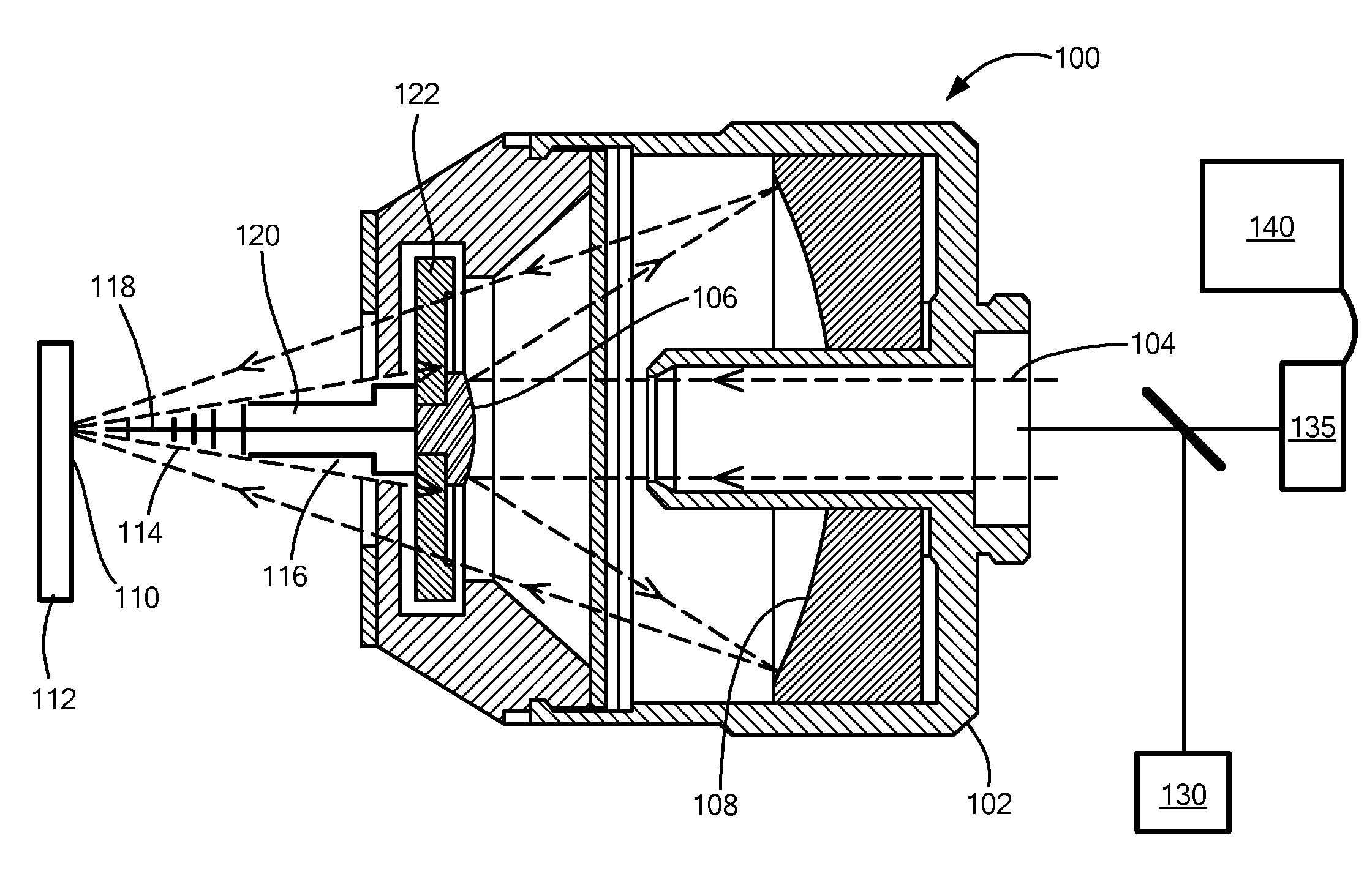
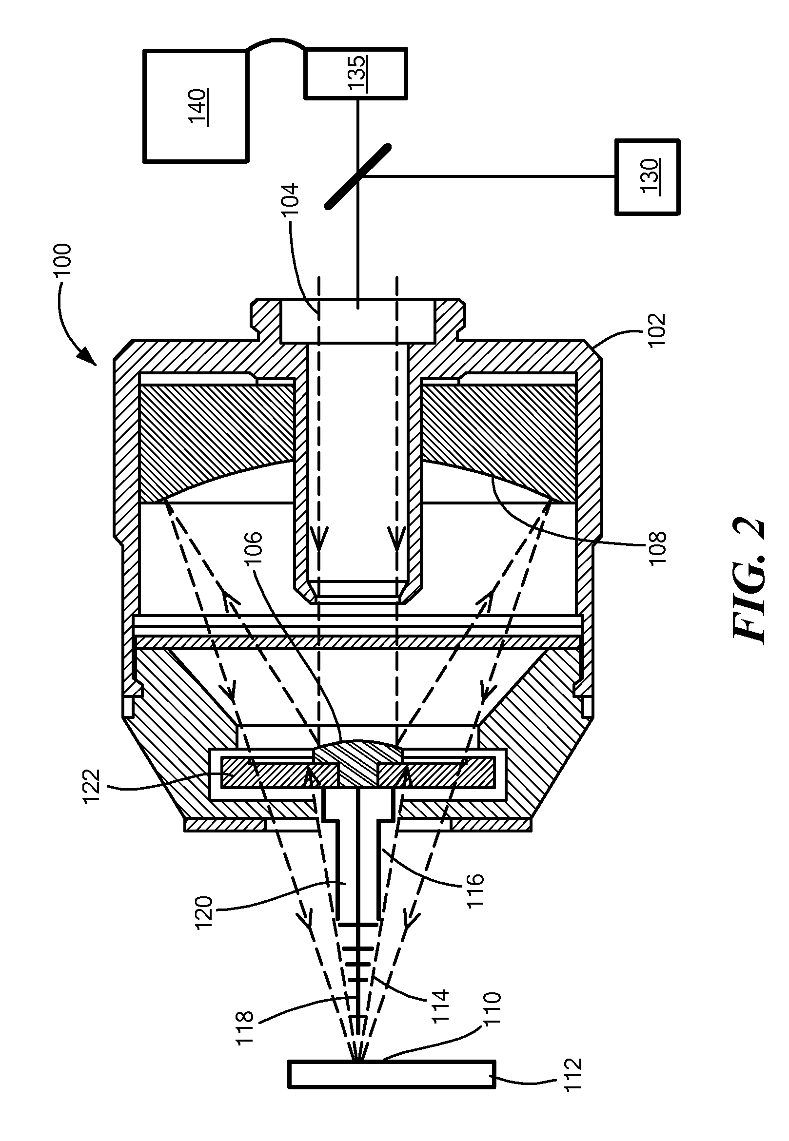
[0008]In accordance with embodiments of the invention, apparatuses and methods are provided for characterizing a sample in situ. In accordance with one set of embodiments of the invention, method are provided having steps of:

[0009]applying a force, by means of a nanoprobe, centered upon a probed locus on the surface of a sample;

[0010]illuminating, with light, a region of the sample including the said probed locus on the surface of the sample;

[0011]measuring a mechanical response of the sample to the applied force; and characterizing an optical interaction between the illuminating light and the region of the sample including the probed locus.

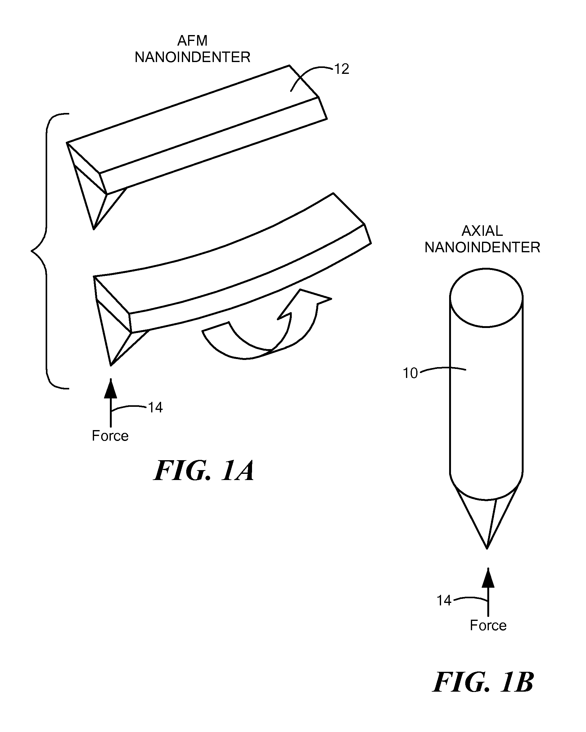
[0012]In accordance with further embodiments of the present invention, the nanoprobe may be a nanoindenter or a tip of an atomic force microscope. The measured mechanical response may be a displacement as a function of force.

[0013]In various alternate embodiments of the present invention, the optical interaction may be light scattering or a non-l...


PUM


Abstract

Methods and apparatus for characterizing a sample in situ as to both its mechanical and optical characteristics. The apparatus comprises a reflective microscope with a concave primary mirror and a convex secondary mirror sharing a common optical axis, and an actuator vignetted by the convex secondary mirror for applying a force to a nanoprobe in a direction having a component along the common optical axis. The apparatus may addition include a source for generating an illuminating beam, a detector, and a processor for forming an image based on a signal provided by the detector.

Description

[0001]The present application claims the priority of U.S. Provisional Patent Application Ser. No. 61 / 915,324, filed Dec. 12, 2013, and incorporated herein by reference.TECHNICAL FIELD[0002]The present invention relates to apparatus and methods for concurrent optical and nanomechanical characterization of a sample.BACKGROUND ART[0003]Mechanobiology studies the effect of physical forces on biological tissue. Directly correlating mechanical and structural information, however, presents a major challenge for both existing imaging and for technologies that provide for characterizing the response of tissue to mechanical force. In the context of mechanobiology, optical microscopy techniques provide noninvasive imaging of biological specimens such as cells, and the extracellular matrix. However, optical imaging methods cannot provide information about the mechanical properties of the imaged tissue. The closest approach to the use of optical techniques to obtain mechanical data has come from...

Claims


Application Information

Patent Timeline
IPC IPC(8): G01L1/24G01Q20/02G02B21/04G01B11/16
CPCG01L1/24G02B21/04G01Q20/02G01B11/16G01Q30/025G01Q60/366
Inventor TOUSSAINT, KIMANIFERREIRA, PLACIDJOHNSON, AMY WAGONERWILSON, WILLIAM
Owner THE BOARD OF TRUSTEES OF THE UNIV OF ILLINOIS
Why Eureka
Try Eureka