
Multiple amplification cycle detection

Active Publication Date: 2015-08-20
BIOFIRE DIAGNOSTICS LLC
View PDF2 Cites 29 Cited by
  • Summary
  • Abstract
  • Description
  • Claims
  • Application Information

AI Technical Summary

Benefits of technology

The present invention provides methods for simultaneously amplifying multiple targets while distinguishing between clinically relevant amplification and other sources of amplification. The methods involve providing a sample with primers for amplifying a locus from each target nucleic acid sequence, subjecting the sample to amplification conditions through multiple amplification cycles, detecting amplification in each cycle, and identifying the target nucleic acid present in the sample. The methods may also include distinguishing between chromosomal integration and clinically-relevant infection by detecting amplification in a sample. Additionally, the invention provides methods for analyzing a target nucleic acid in a sample by amplifying it and detecting it using a melt curve analysis. The methods may also involve generating a corrected amplification curve for the target nucleic acid and plotting it. The invention also includes reaction vessels and devices for carrying out the methods.

Problems solved by technology

Traditional microbiology techniques for diagnosing pathogens can take days or weeks, often delaying a proper course of treatment.
A challenge to using PCR as a primary means of diagnosis is the variety of possible causative organisms and the low levels of organism present in some pathological specimens.
It is often impractical to run large panels of PCR assays, one for each possible causative organism, most of which are expected to be negative.
The problem is exacerbated when pathogen nucleic acid is at low concentration and requires a large volume of sample to gather adequate reaction templates.
In some cases, there is inadequate sample to assay for all possible etiological agents.
While multiplex PCR has proven to be valuable in some systems, shortcomings exist concerning robustness of high level multiplex reactions and difficulties for clear analysis of multiple products.
However, this further handling can be expensive and may lead to contamination or other problems.
While such environmental contamination tends to be present in concentrations that are significantly below that of a clinically relevant sample, it can be difficult to distinguish between environmental contamination and clinical infection.

Method used


Image

  • Multiple amplification cycle detection
  • Multiple amplification cycle detection
  • Multiple amplification cycle detection

Examples


example 1

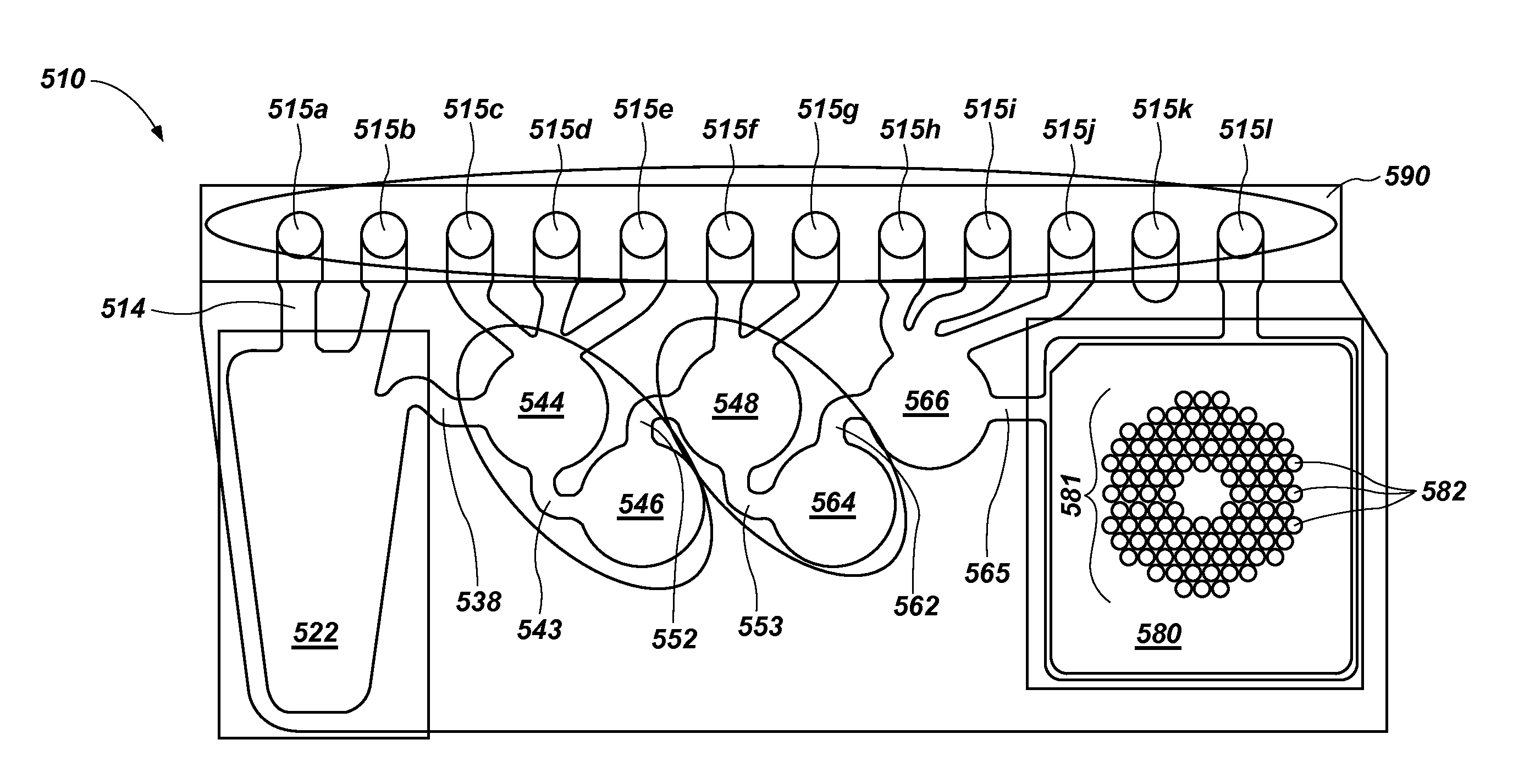
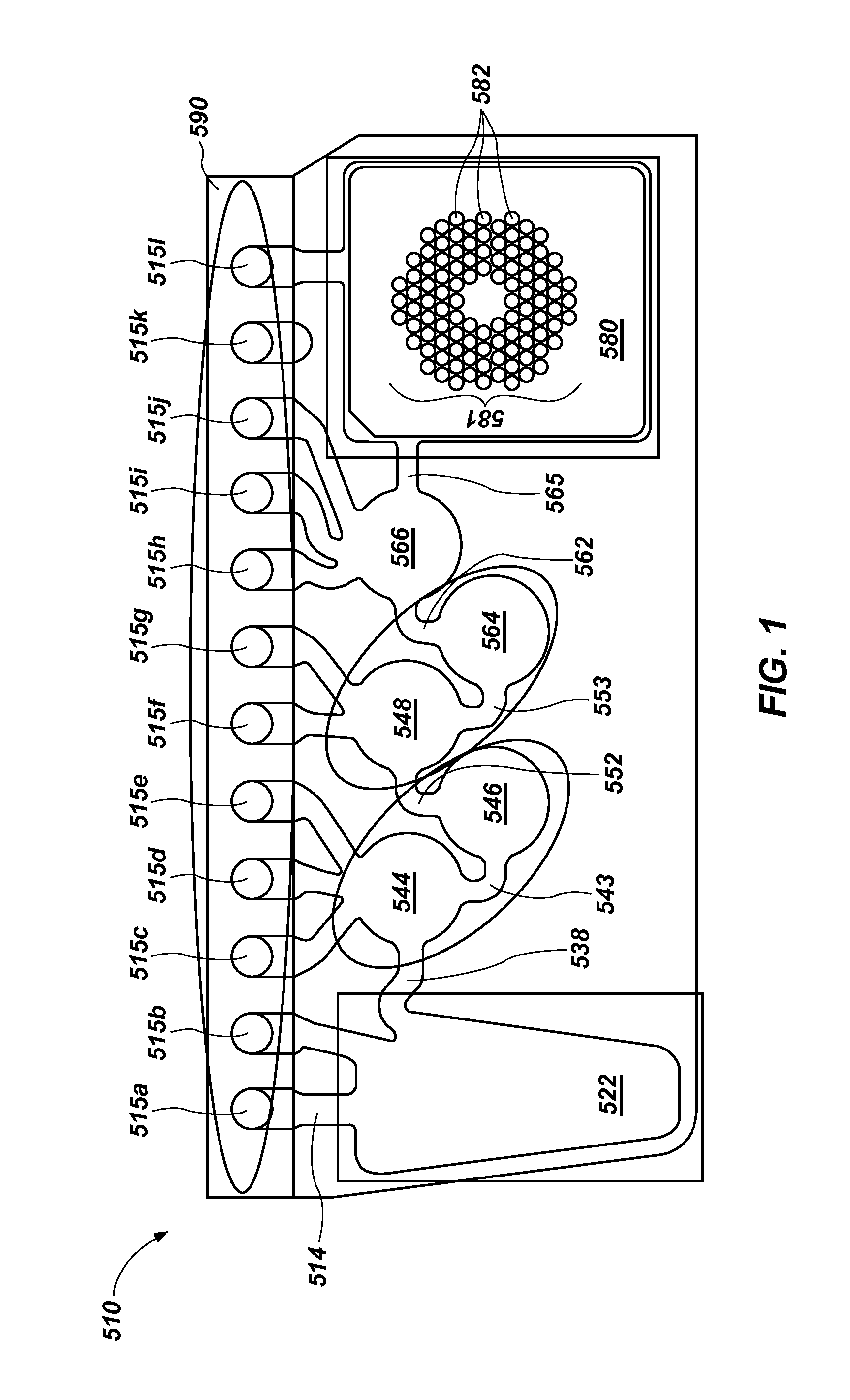
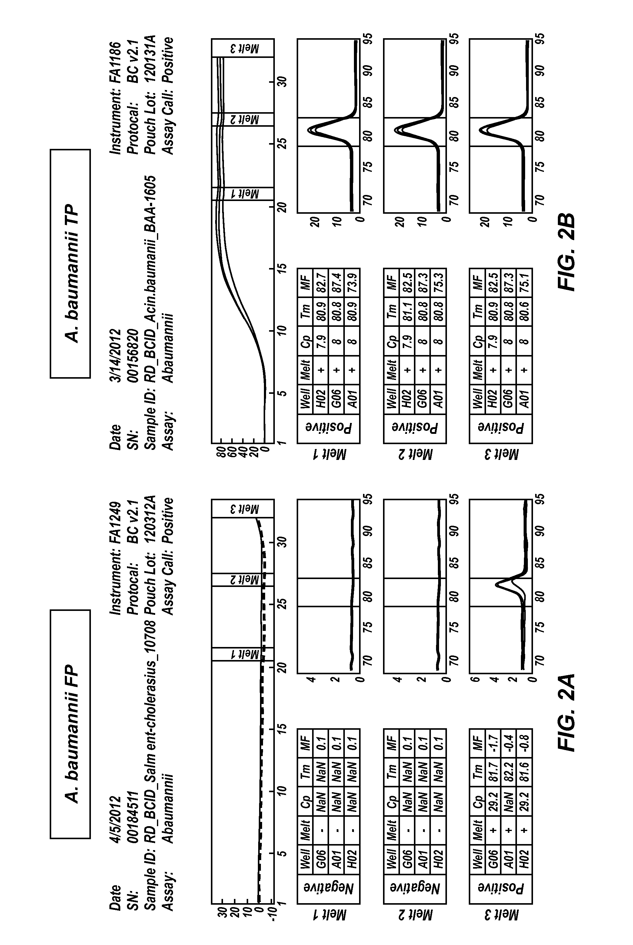
[0058]The FilmArray Blood Culture Identification (BCID) system is designed to provide rapid identification of a broad range of microorganism pathogens directly from blood culture. The illustrative BCID panel detects the most common bacteria and yeast isolated from positive aerobic blood cultures (PABC), as well as select antibiotic resistance genes, with ≧95% sensitivity. A commercial BCID panel is available from BioFire Diagnostics, Inc. This example uses a research version of the FilmArray BCID panel to demonstrate methods of distinguishing between true positives and environmental contamination.

[0059]Various gram-positive and gram-negative bacteria, as well as Candida yeast isolates were tested for assay reactivity. Mock PABC samples were prepared by spiking microorganism into a mixture of human whole blood and BD BACTEC Aerobic Plus / F blood culture medium. Microorganisms were spiked at concentrations consistent with that observed for blood culture bottles that had recently been i...

example 2

[0072]In Example 1, melts at different cycle numbers were used to distinguish between environmental contamination and clinical infection, wherein each test in the panel was assigned a cycle number, and positives and negatives were called based on the result at the assigned cycle number. Using different cycle numbers for calls can also be used to distinguish between potential “false positives” where nucleic acid is present at substantial quantities but not clinically relevant and clinically relevant true positives that do not have a crossing point until a later cycle. One such example is with latent viral infection through chromosomal integration, wherein the chromosomally integrated viral DNA may or may not be responsible for the clinical symptoms.

[0073]For example, an individual may have inherited the HHV6 virus from a parent who had been infected with the virus and the virus was latently chromosomally integrated (termed chromosomally-integrated HHV6, “ciHHV6”). This individual wou...

example 3

[0075]In Examples 1 and 2, different cycle numbers were used to distinguish between environmental contamination, potentially non-clinically relevant infection, and clinically-relevant infection. In this example, additional cycles are used to enable detection of low level true positives. In this method, the detection and identification method is a modified two-step process. The first step is a set amplification protocol, optionally with additional melt cycles as used in Examples 1 and 2, and the second step employs a higher signal-to-noise detection during at least one subsequent melt. An illustrative protocol is shown in FIG. 5.

[0076]As shown in FIG. 5, a set number of amplification cycles, illustratively 26 cycles, are run. Any wells that return a positive result at cycle 26 optionally need not be analyzed further. The positive result may be made by amplification curve, or may be made or confirmed by a melt curve analysis as discussed above, for those samples that show amplificatio...


PUM

PropertyMeasurementUnit
Temperatureaaaaaaaaaa
Poweraaaaaaaaaa
Concentrationaaaaaaaaaa

Abstract

Methods and devices are provided for simultaneously amplifying a plurality of sample wells for a predetermined amount of amplification, detecting whether amplification has occurred in a first set of the wells, amplifying for an additional amount of amplification and detecting whether amplification has occurred in a second set of the wells. Methods are also provided for analyzing a target nucleic acid sequence using melt curves that were generated in a plurality of amplification cycles.

Description

CROSS-REFERENCE TO RELATED APPLICATION[0001]This application claims the benefit of and priority to U.S. Provisional Patent Application No. 61 / 699,103, filed on Sep. 10, 2012, entitled “Multiple Amplification Cycle Detection”, the entirety of which is incorporated herein by reference.GOVERNMENT INTEREST[0002]This invention was made with government support under Grant No. 1U01 AI082184 awarded by National Institutes of Health. The government has certain rights in the invention.BACKGROUND OF THE INVENTION[0003]In the United States, Canada, and Western Europe infectious disease accounts for approximately 7% of human mortality, while in developing regions infectious disease accounts for over 40% of human mortality. Infectious diseases lead to a variety of clinical manifestations. Among common overt manifestations are fever, pneumonia, meningitis, diarrhea, and diarrhea containing blood. While the physical manifestations suggest some pathogens and eliminate others as the etiological agent...

Claims


Application Information

Patent Timeline
IPC IPC(8): C12Q1/68
CPCC12Q1/686C12Q2537/143C12Q2527/107C12Q1/6851C12Q1/689C12Q1/705C12Q2537/165
Inventor RASMUSSEN, RANDY P.CRISP, ROBERT JOHNHEMMERT, ANDREW CLINTONCAMPBELL, ELIZABETH BARKERROBBINS, THOMAS CHARLESEYRE, DAVID J.
Owner BIOFIRE DIAGNOSTICS LLC
Learn More