Photodynamic diagnosis apparatus provided with collimator

a technology of collimator and diagnosis apparatus, which is applied in the field of photodynamic diagnosis apparatus provided with collimator, can solve the problems of weakening fluorescence emission, difficult operation, and failure to output excitation light, and achieve the effect of efficient light collection

Inactive Publication Date: 2015-08-27
SBI PHARMA CO LTD
View PDF6 Cites 12 Cited by
  • Summary
  • Abstract
  • Description
  • Claims
  • Application Information

AI Technical Summary

Benefits of technology

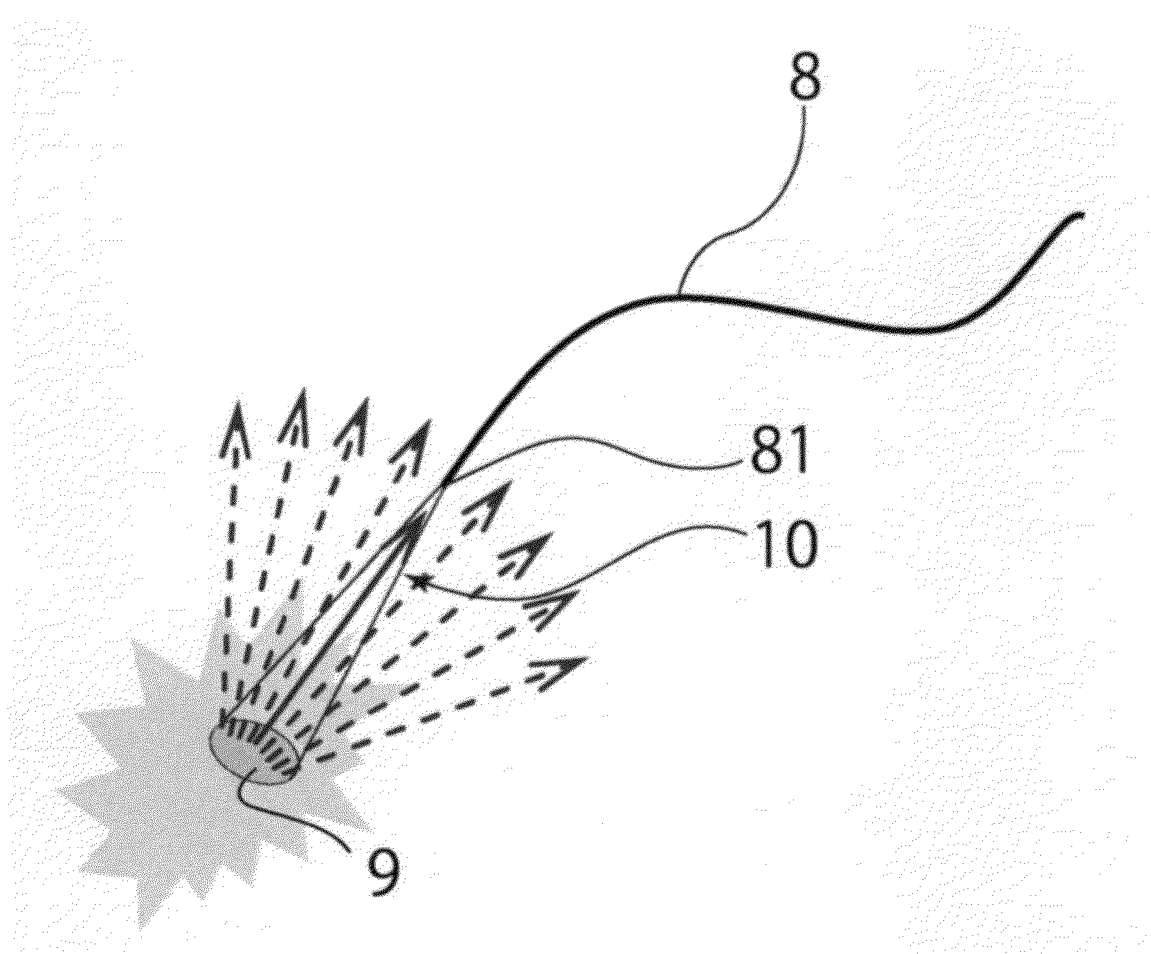
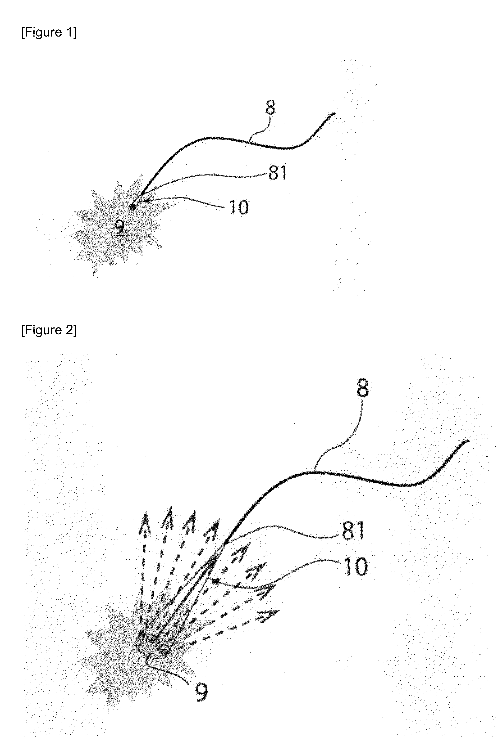
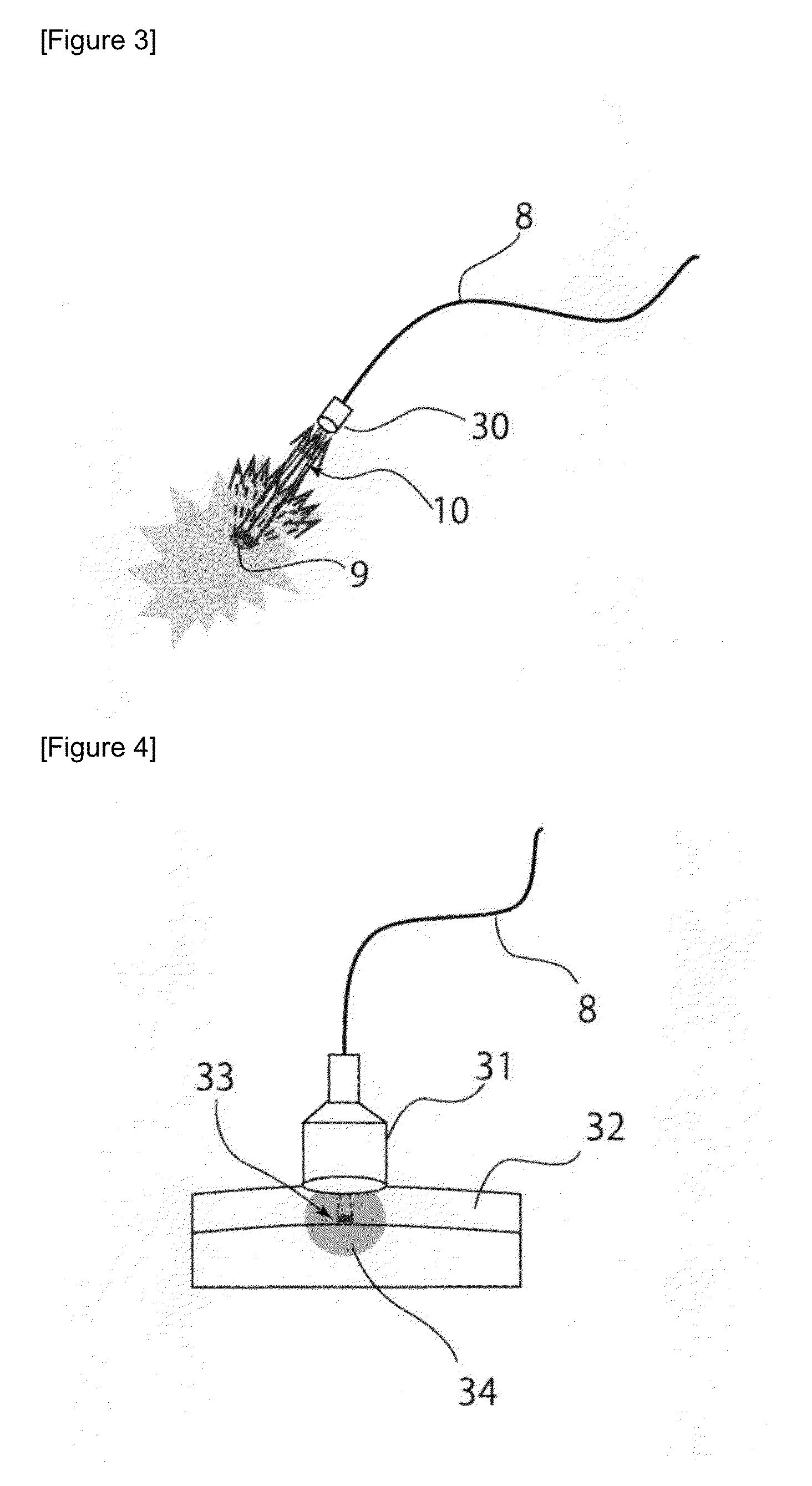
[0022]A diagnosis apparatus according to the present invention including an excitation light irradiation/fluorescence detection optical fiber with a collimator connected to an output/input end thereof enables efficient light collection by making not only excitation light but also fluorescence produced by an excited photosensitizing substance pass through the collimator. As a result, even if there is a distance from an irradiation portion of the optical fiber to a diseased tissue, a fluorescence detection sensitivity equivalent to that in a case where irradiation is provided with the irradiation portion of the optical fiber brought close to the diseased tissue can be obtained. For example, use of a surgical microscope having a fluorescence diagnosis mode, in which an excitation light irradiation/fluorescence detection optical fib...

Problems solved by technology

Thus, during irradiation with violet laser, if a distal end of an optical fiber comes into contact with a tissue, the distal end largely absorbs, in particular, hemoglobin in the blood, which sticks to or is carbonized on an end face, which may result in failure to output excitation light, and in each of such cases, a surgeon needs to clean the distal end with an alcohol swab.
Also, in intraoperative fluorescence diagnosis for brain tumor, which is widely used in clinical studies, an operation is performed with a suction tube in one hand and a bipolar or ultrasound surgical knife or the like in the other hand, and thus, if it is ne...

Method used


Image

  • Photodynamic diagnosis apparatus provided with collimator
  • Photodynamic diagnosis apparatus provided with collimator
  • Photodynamic diagnosis apparatus provided with collimator

Examples


example 1

1. Configuration

[0067]Specifications of an intraoperative diagnosis system including a combination of the diagnosis apparatus according to the present invention illustrated in FIG. 7 and a surgical microscope used for brain tumor removal surgery are indicated below.

Detection method: sound in PpIX detection for detecting a peak of a spectrum of PpIX fluorescence derived from a violet laser diode; the pulse sound interval is changed when the PpIX fluorescence intensity increases.

Excitation light irradiation / fluorescence detection optical fiber: single core fiber (core diameter: φ300 μm) Collimator: F220SMA-A manufactured by Thorlabs, Inc.

Laser type / wavelength: InGaN laser diode / 406 nm (typical)

Laser output / class: fiber output 70 mW (CW operation) / class 3B

Surgical microscope: “OPMI Pentero” manufactured by Carl Zeiss

[0068]FIG. 8 is a fluorescence diagnosis mode-provided surgical microscope having a function that provides violet illuminating light to an operative field by inserting / retr...


PUM


Abstract

An object of the present invention is to provide a PDD diagnosis apparatus that even if there is a distance from an irradiation portion of an optical fiber to a diseased tissue, enables provision of a fluorescence detection sensitivity equivalent to that in a case where irradiation is provided in such a manner that the irradiation portion of the optical fiber is brought close to the diseased tissue. A diagnosis apparatus including: a light source unit that emits excitation light to a photosensitizing substance accumulated in a diseased tissue; a detection unit that detects fluorescence produced from the photosensitizing substance; and an excitation light irradiation/fluorescence detection optical fiber including an output/input end with a collimator connected thereto, in which the excitation light emitted from the light source unit is guided to the excitation light irradiation/fluorescence detection optical fiber, and the excitation light output from the output/input end is provided in such a manner that the excitation light is collimated by the collimator and excites the photosensitizing substance accumulated in the diseased tissue; and the fluorescence produced by the photosensitizing substance excited by the excitation light is input from the output/input end via the collimator, and the input fluorescence is guided to the excitation light irradiation/fluorescence detection optical fiber and the fluorescence produced from the photosensitizing substance is detected in the detection unit is used.

Description

TECHNICAL FIELD[0001]The present invention relates to a photodynamic diagnosis apparatus provided with a collimator, and more specifically relates to a diagnosis apparatus for identifying a diseased tissue (focal site), the apparatus including, e.g., a light source unit that emits excitation light, a detection unit that detects fluorescence produced from a photosensitizing substance, and an excitation light irradiation / fluorescence detection optical fiber including an output / input end with a collimator connected thereto.BACKGROUND ART[0002]Cancer (malignant tumor) is the greatest killer in Japan, and one out of every two Japanese persons is said to be affected by cancer. In the other developed countries, cancer is also high in the list of death causes. Therefore, development of effective diagnostic methods and therapeutic methods for cancers is the earnest wish of people of the world including Japanese people.[0003]As a cancer diagnostic method, photodynamic diagnosis (hereinafter r...

Claims


Application Information

Patent Timeline
IPC IPC(8): A61B5/00A61B1/07A61B1/06
CPCA61B5/0071A61B5/0084A61B5/7405A61B2562/0238A61B1/063A61B1/07A61B5/7282A61B1/00096A61B1/043G01N21/645G01N2021/6484G02B23/26A61B1/00167A61B1/0607G02B21/0012G02B23/2469
Inventor INOUE, KATSUSHIHIRAMITSU, MASANORINAKAJIMA, MOTOWOTANAKA, TOHRU
Owner SBI PHARMA CO LTD
Why Eureka
Try Eureka