Arthroscopic joint surgery device and method of use

a technology for arthroscopic joints and obturators, which is applied in the field of arthroscopic trocars or obturators for joint surgery, can solve the problems of affecting the treatment effect of patients, affecting the treatment effect, so as to achieve the effect of less trauma and pain and easy handling

Inactive Publication Date: 2015-12-17
NEEDLEMAN RICHARD L
View PDF4 Cites 0 Cited by
  • Summary
  • Abstract
  • Description
  • Claims
  • Application Information

AI Technical Summary

Benefits of technology

[0007]In one embodiment, the knob can be truncated and ridged for easy handling by a surgeon.
[0008]For the purposes of this disclosure, an angiocatheter is a device that can be inserted into a blood vessel, usually a vein. Medications, hydrating fluid, and / or products such as blood can be administered through this access port into the blood system. A metal stylet is within a cannulated flexible plastic tube. The metal stylet has a sharp point to allow for penetration of the skin and soft structures between the skin and the vein. It also allows for penetration into the vein. It is inserted into a vein with the metal stylet in to keep it rigid. This is important during insertion. Once the catheter is within the vein, the stylet is removed (and the plastic catheter is advanced further into the vein) so that medications, hydrating fluid, and / or products such as blood can be administered through this access port into the blood system. Meanwhile, the catheter becomes flexible that allows for less trauma and pain while it remains within the blood vessel.
[0009]For the purposes of this disclosure, a cannulated needle is a device that can be inserted near the spinal cord. It is called a ‘spinal needle’. Medication can be administered or a specimen of spinal fluid can be obtained through this access port. A metal stylet is within a rigid metal tube. The metal stylet has a sharp point to allow for penetration of the skin and soft structures between the skin and the spinal cord. It is inserted near the spinal cord with the metal stylet in. Once the needle is in the proper position next to the spinal cord, the stylet is removed so that medication can be administered or a specimen of spinal fluid can be obtained through this access port. The needle remains rigid which allows for it to remain in the appropriate position in the short time that the procedure is performed.

Problems solved by technology

The hip had been a difficult joint to arthroscope because it is a deep joint with a thick layer of overlying soft tissues that were not amenable to palpating external anatomic landmarks accurately, especially in obese patients.
The sharp-ended trocar can scuff and damage a patient's joint cartilage.
However, a blunt-ended obturator may not be able to penetrate a thick joint capsule.

Method used


Image

  • Arthroscopic joint surgery device and method of use
  • Arthroscopic joint surgery device and method of use
  • Arthroscopic joint surgery device and method of use

Examples


Embodiment Construction

[0034]The present disclosure discusses an arthroscopic joint surgery device and method of use. The device and method of use enable improved surgery techniques in medium and small joints such as ankle joints. Previously, these improved surgery techniques were only available in larger joints such as the hip joint.

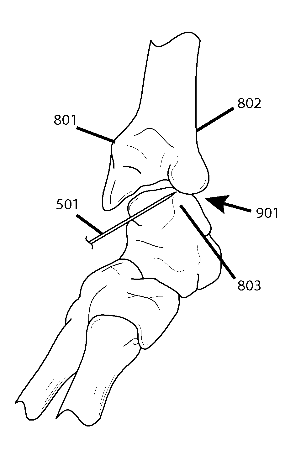
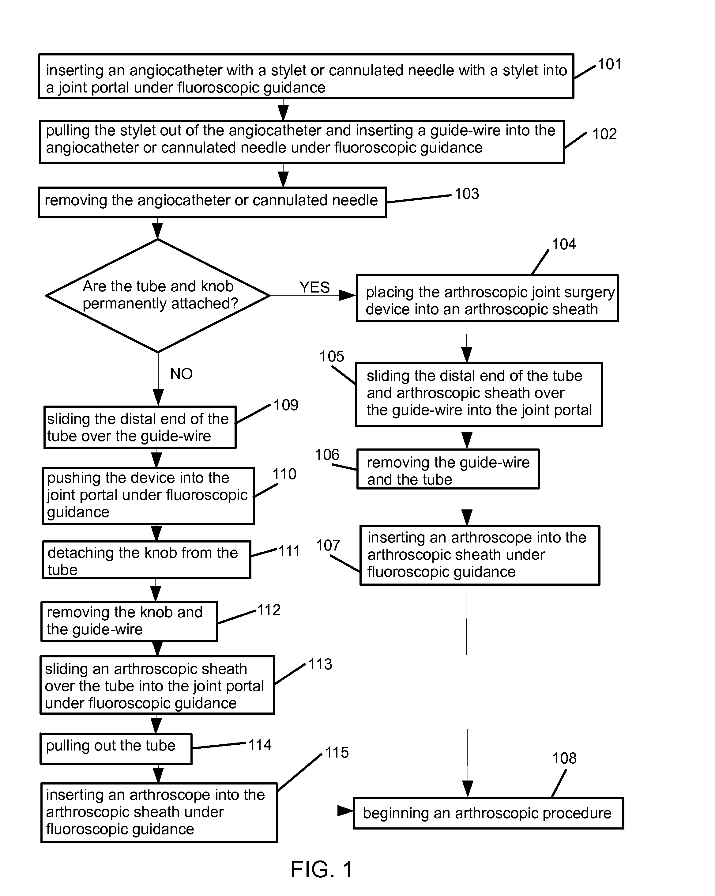
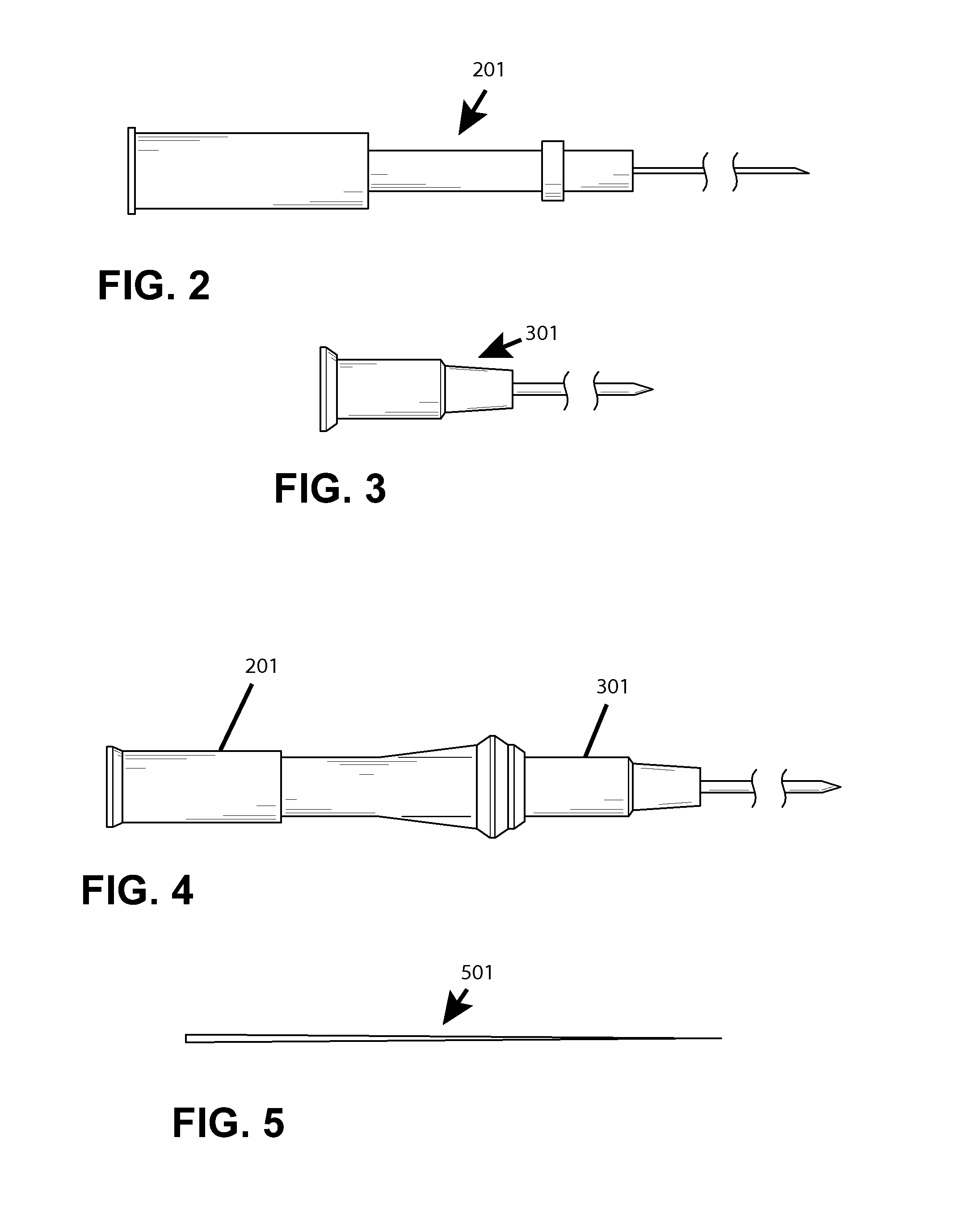
[0035]The method is an adaptation of common hip surgery techniques to small joint arthroscopy by performing the procedure under the guidance of fluoroscopy, placing an angiocatheter with a stylet or a cannulated needle with a stylet into the joint portal prior to dilation, placing a blunt guide-wire into the angiocatheter or cannulated needle, and using the device as described herein to dilate the joint portal opening prior to inserting an arthroscopic sheath.

[0036]The soft issues overlying the anterior ankle are thin and the anatomic landmarks for portal placement are usually palpable. Oftentimes under pathologic conditions, the ankle joint is swollen with edema and conseque...


PUM


Abstract

An arthroscopic joint surgery device and method of use. The device has a tube and a knob with a hole that has an inner diameter matching the inner diameter of the tube and mates with the inner diameter of the tube. The tube and knob can be permanently attached or the knob can be configured to enable the knob to be unattached from the tube. The utilization of the device in this method enables surgeons to safely and efficiently locate the joint portals for the placement of the arthroscope and for a separate inflow portal. The arthroscope is in the proper position to visualize the whole joint. Damage to the joint surface cartilage is minimized. After needle localization of the joint under fluoroscopic guidance, the method uses the device to dilate a joint portal opening, such as an anteromedial ankle portal in preparation for the placement of the arthroscope or the posterolateral portal for the introduction of the inflow cannula. The method is performed under fluoroscopic guidance.

Description

TECHNICAL FIELD[0001]The present disclosure relates to the field of arthroscopic trocar or obturator devices for joint surgery and their methods of use. More particularly, the disclosure discusses devices and methods for smaller joints such as an ankle.BACKGROUND[0002]Trocar and obturator devices are currently successfully used for arthroscopic knee and shoulder surgery. These joints have a thin layer of overlying soft tissue so that the surgeon can identify the external anatomic landmarks and develop small portals into the joints. The hip had been a difficult joint to arthroscope because it is a deep joint with a thick layer of overlying soft tissues that were not amenable to palpating external anatomic landmarks accurately, especially in obese patients. Recently, there has been a development of arthroscopic hip surgery by using external anatomic topography, noninvasive distraction, and fluoroscopy. Modifications have been made to the basic knee and shoulder arthroscopic instrument...

Claims


Application Information

Patent Timeline
Patent Type & Authority Applications(United States)
IPC IPC(8): A61B17/34A61B17/00
CPCA61B17/3423A61B2017/3456A61B17/3415A61B17/00234A61B17/3401A61B17/3421A61B2017/22038A61B2090/376
Inventor NEEDLEMAN, RICHARD L.
Owner NEEDLEMAN RICHARD L
Why Eureka
Try Eureka