Functional Magnetic Resonance Imaging (fMRI) Methodology Using Transverse Relaxation Preparation and Non-Echo-Planar Imaging (EPI) Pulse Sequences

a magnetic resonance imaging and pulse sequence technology, applied in the field of magnetic resonance imaging, can solve the problems of geometric distortion and signal dropout, scale with the square of field strength, limit the acquisition efficiency and temporal resolution of fmri, etc., to suppress inflow effects, short echo time, and low-high (centric) phase encoding

Inactive Publication Date: 2016-04-28
THE JOHN HOPKINS UNIV SCHOOL OF MEDICINE +1
View PDF7 Cites 11 Cited by
  • Summary
  • Abstract
  • Description
  • Claims
  • Application Information

AI Technical Summary

Benefits of technology

[0008]In accordance with an aspect of the present invention, the method includes providing the single-shot fast-GRE readout having a short echo time and using the short echo time of approximately <2 ms. The method includes using the single shot fast-GRE readout taking the form of at least one of turbo field echo, TFE, or turbo flash. The method includes acquiring the image in the form of a whole brain fMRI image with minimal distortion and dropouts and acquiring the image with a spatial resolution of approximately mm isotropic. Additionally, the method includes acquiring the image having a temporal resolution of 2.3 s at 7 T. The BOLD contrast is generated before providing the single-shot, fast-gradient echo (GRE) readout. Two 180° pulses in the T2-weighted preparation module can be used to compensate for phase variations and to suppress inflow effects. A spoiler gradient can be played at an end of the T2-weighted preparation module on a first phase encoding axis that has a lowest gradient duty cycle to dephase any residual transverse magnetization. A SINC RF pulse can be used for refocusing, and the single-shot fast-gradient echo readout can have low-high (centric) phase encoding.
[0011]In accordance with yet another aspect of the present invention, the steps include providing the single-shot fast-GRE readout having a short echo time and using the short echo time of approximately <2 ms. The steps include using the single shot fast-GRE readout taking the form of at least one of turbo field echo, TFE, or turbo flash. The steps include acquiring the image in the form of a whole brain fMRI image with minimal distortion and dropouts and acquiring the image with a spatial resolution of approximately 2.5 mm isotropic. Additionally, the steps include acquiring the image having a temporal resolution of 2.3 s at 7 T. The BOLD contrast is generated before providing the single-shot, fast-gradient echo (GRE) readout. Two 180° pulses in the T2-weighted preparation module can be used to compensate for phase variations and to suppress inflow effects. A spoiler gradient can be played at an end of the T2-weighted preparation module on a first phase encoding axis that has a lowest gradient duty cycle to dephase any residual transverse magnetization. A SNC RF pulse can be used for refocusing, and the single-shot fast-gradient echo readout can have low-high (centric) phase encoding.

Problems solved by technology

While they provide excellent sensitivity to signal changes during functional stimulation with high acquisition efficiency, they often suffer from geometric distortions and signal dropouts in regions near air cavities such as the orbitofrontal cortex and temporal lobes, which are exacerbated at high field.
One of the main constraints for SE sequences, however, is the high power deposition imposed mainly by the large number of refocusing radiofrequency (RF) pulses, which unfortunately scales with the square of the field strength.
More importantly, the T2 or T2* contrast in most BOLD fMRI methods is generated during the imaging sequence, which may impose some intrinsic constraints.
For instance, a long echo time (TE) is required for SE BOLD, which produces some “dead time” that limits the acquisition efficiency and temporal resolution for fMRI.

Method used


Image

  • Functional Magnetic Resonance Imaging (fMRI) Methodology Using Transverse Relaxation Preparation and Non-Echo-Planar Imaging (EPI) Pulse Sequences
  • Functional Magnetic Resonance Imaging (fMRI) Methodology Using Transverse Relaxation Preparation and Non-Echo-Planar Imaging (EPI) Pulse Sequences
  • Functional Magnetic Resonance Imaging (fMRI) Methodology Using Transverse Relaxation Preparation and Non-Echo-Planar Imaging (EPI) Pulse Sequences

Examples


example

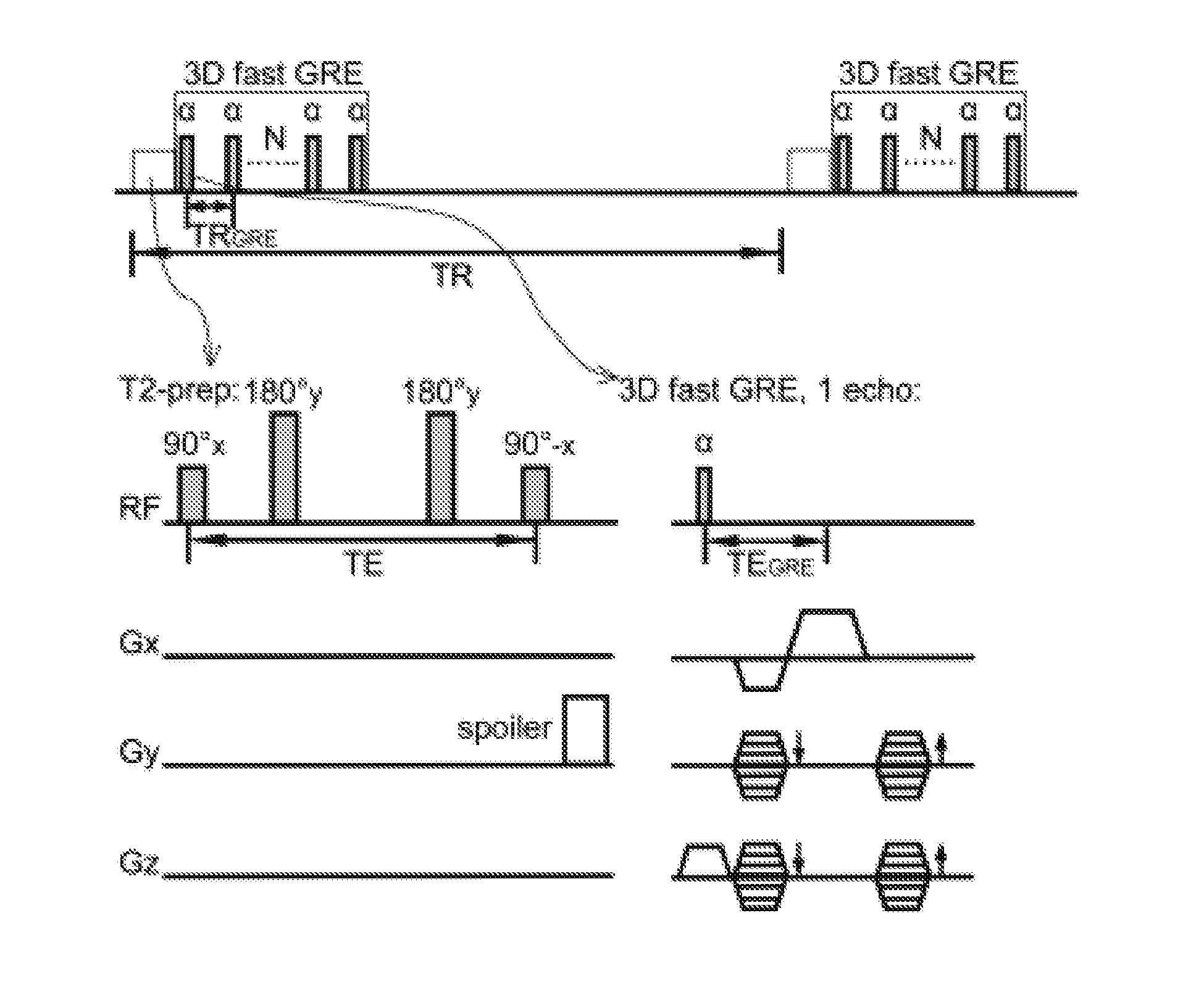
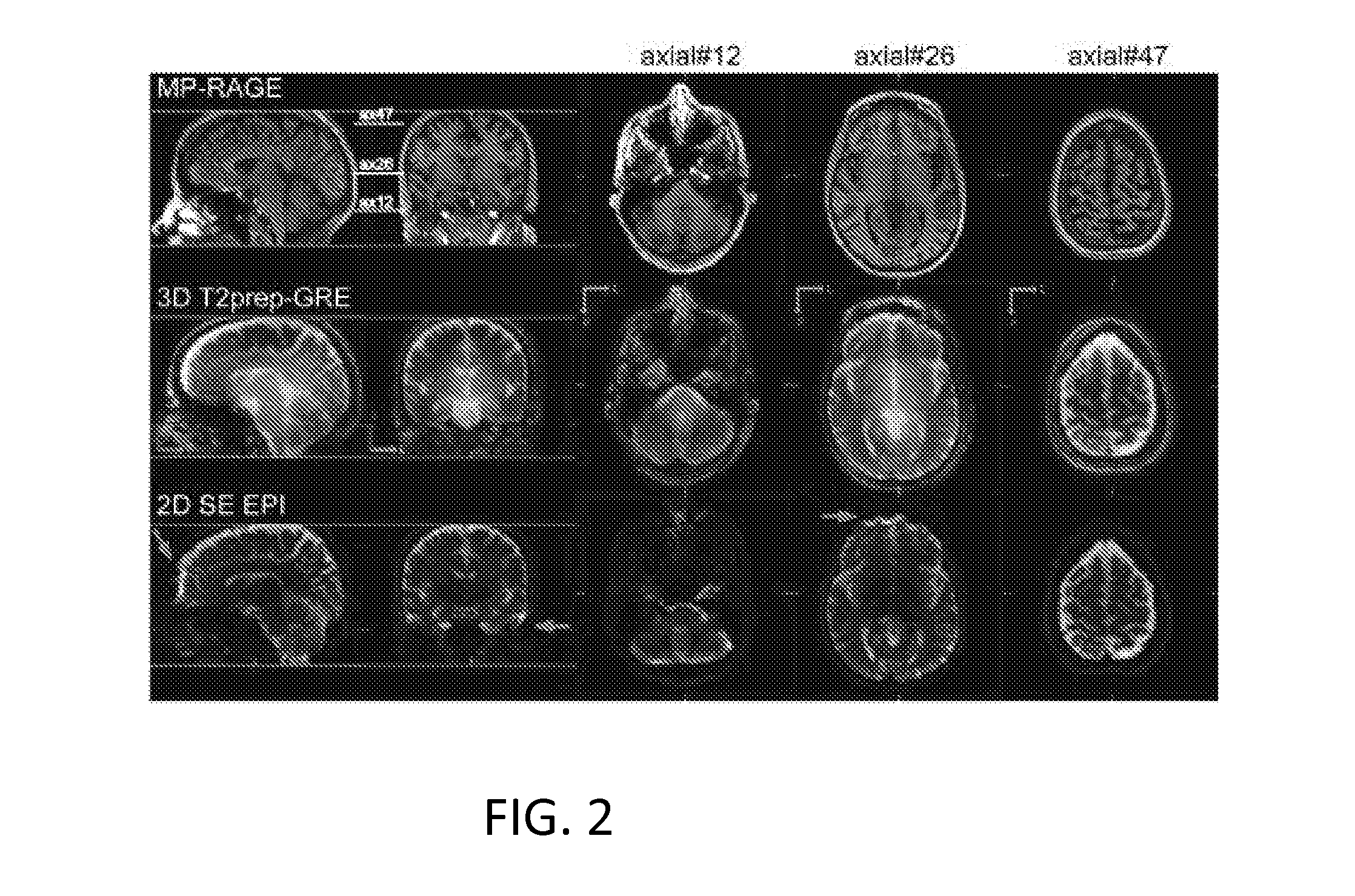
[0032]An exemplary implementation of the present invention is described herein, in order to further illustrate the present invention. The exemplary implementation is included merely as an example and is not meant to be considered limiting. Any implementation of the present invention on any suitable subject known to or conceivable by one of skill in the art could also be used, and is considered within the scope of this application.

[0033]Five healthy human subjects, who gave written informed consent before participating in this Johns Hopkins Institutional Review Board (IRB) approved, Health Insurance Portability and Accountability Act (HIPAA)-compliant study, were scanned on a 7 T Philips MRI scanner (Philips Healthcare, Best, The Netherlands). A 32-channel phased-array head coil (Nova Medical, Wilmington, Mass.) was used for RF reception and a head-only quadrature coil for transmit. Two rectangular pads (23×10×2 mm) filled with high dielectric constant materials were placed between t...


PUM


Abstract

An embodiment in accordance with the present invention provides a new acquisition scheme for T2-weighted BOLD fMRI. It employs a T2 preparation module to induce the BOLD contrast, followed by a single-shot 3D fast gradient echo (GRE) readout with short echo time (TE<2 ms). The separation of BOLD contrast generation from the readout substantially reduces the “dead time” due to long TE required in spin echo (SE) BOLD sequences. This approach termed “3D T2prep-GRE,” can be implemented with any magnetic resonance imaging machine, known to or conceivable by one of skill in the art. This approach is expected to be useful for ultra-high field fMRI studies that require whole brain coverage, or focus on regions near air cavities. The concept of using T2 preparation to generate BOLD contrast can be combined with many other fast imaging sequences at any field strength.

Description

CROSS REFERENCE TO RELATED APPLICATIONS[0001]This application claims the benefit of U.S. Provisional Patent Application No. 61 / 830,360 filed on Jun. 3, 2013, which is incorporated by reference, herein, in its entirety.GOVERNMENT SUPPORT[0002]This invention was made with government support under NIH RO15P41-RR015241 awarded by the National Institutes of Health. The government has certain rights in the invention.FIELD OF THE INVENTION[0003]The present invention relates generally to imaging. More particularly the present invention relates to a system and method for magnetic resonance imaging.BACKGROUND OF THE INVENTION[0004]High field [7 Tesla (T)] human MRI scanners have become available in recent years with the promise of an approximately linear increase in signal-to-noise ratio (SNR) with field strength. In addition, high field is particularly attractive to blood-oxygenation-level-dependent (BOLD) functional MRI (fMRI) as the BOLD contrast shows a supra-linear increase with field st...

Claims


Application Information

Patent Timeline
Patent Type & Authority Applications(United States)
IPC IPC(8): A61B5/00
CPCA61B2576/026A61B5/0042A61B5/055G01R33/4806G01R33/5602G01R33/5617G16H30/40
Inventor HUA, JUNJONES, CRAIG KENNETHQIN, QINVANZIJL, PETER
Owner THE JOHN HOPKINS UNIV SCHOOL OF MEDICINE
Why Eureka
Try Eureka