
Systems, devices, apparatus and method devices for providing endoscopic mucosal therapy

Inactive Publication Date: 2016-07-07
THE GENERAL HOSPITAL CORP
View PDF1 Cites 0 Cited by
  • Summary
  • Abstract
  • Description
  • Claims
  • Application Information

AI Technical Summary

Benefits of technology

The patent is about a new system that can treat tissue abnormalities in the body, such as those in the esophagus, lung, or gastrointestinal organs. Unlike other treatment methods, this system uses a therapeutic effect to alter the shape of the tissue abnormality, rather than surgically removing it. The treatment can involve various methods such as phototherapy or conductive heating to damage the tissue. This can help to reduce damage to the surrounding tissue and can even allow the tissue to remain in place for a limited time after the treatment. The system is designed to minimize the risk of complications and can be used with an endoscope or other minimally invasive procedures. The treatment can be repeated if necessary.

Problems solved by technology

Because of the radio-frequency field spatial confinement, the damage is limited to the tissues adjacent to the device.
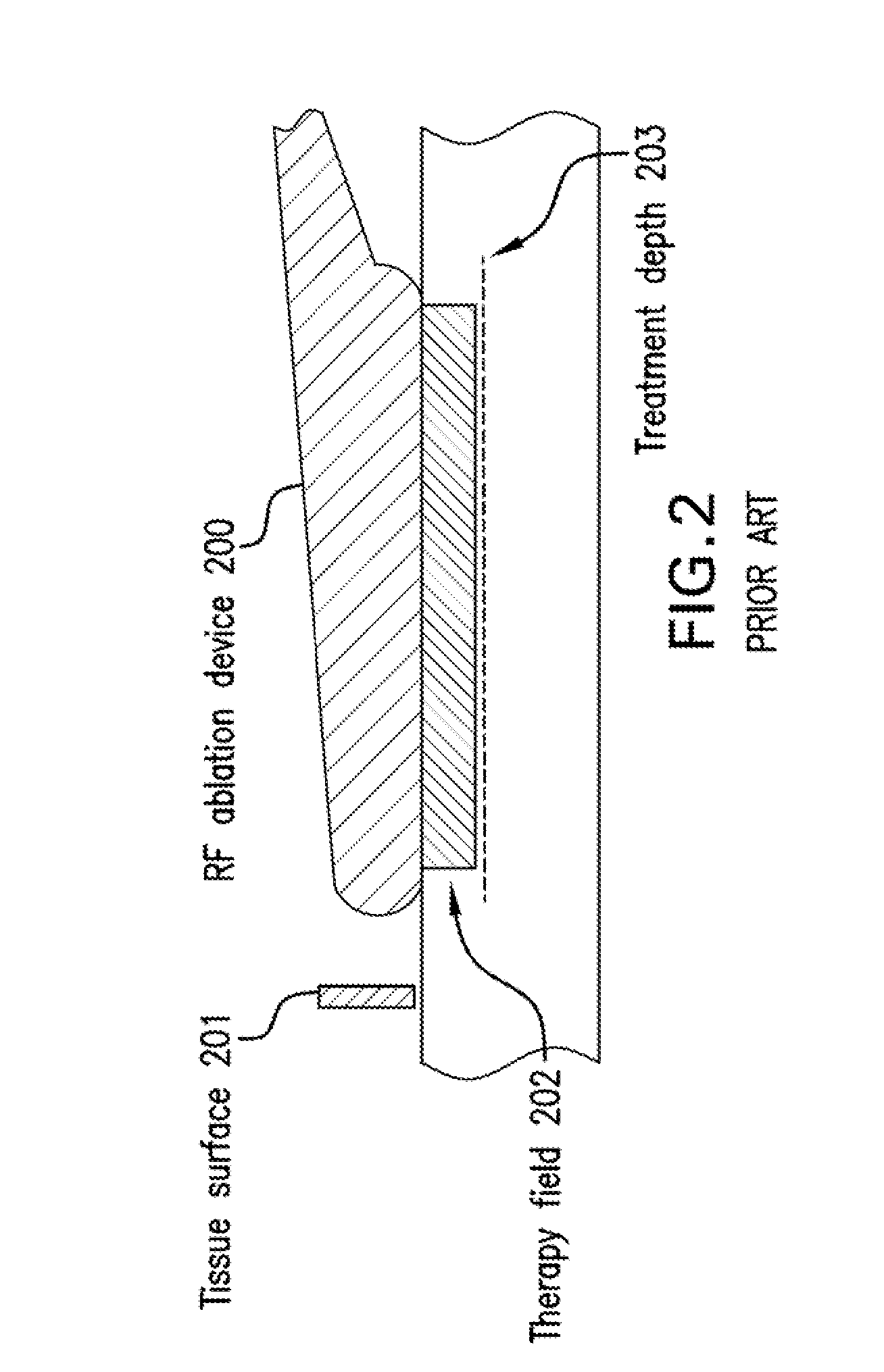
One challenge associated with existing endoscopic mucosal therapy deployments can be that the depth of the therapy is not intrinsically limited to anatomical boundaries, but instead is typically induced to a fixed distance from the device at the time of therapy.
For these reasons, among others, existing methods and system for providing an endoscopic mucosal therapy can require multiple treatments sessions to eliminate the lesion, or can be result in damage to tissue anatomical layers that are not intended to be targeted.
Further, EMR resects tissue leaving some bleeding and scarring.

Method used


Image

  • Systems, devices, apparatus and method devices for providing endoscopic mucosal therapy
  • Systems, devices, apparatus and method devices for providing endoscopic mucosal therapy
  • Systems, devices, apparatus and method devices for providing endoscopic mucosal therapy

Examples


embodiment

For Integration with Endoscopes and Large Area Treatment

[0051]Various exemplary methods or devices to endoscopically deploy these exemplary therapy devices can be used. According to exemplary embodiments of the configurations, systems, apparatus, devices and / or designs of the present disclosure illustrated in FIG. 14, the therapy device 1400 can be affixed or otherwise connected to an endoscope 1401 using a mounting 1402, and can hang as a paddle that occupies a portion of the endoscopes' circumferential view, for example 90 degrees. This exemplary paddle can extend into the endoscope's imaging field, as shown in FIG. 14, such that the treated tissue can be visualized, or can be drawn proximally to move it outside the imaging field, or alternatively can be designed or configured to move dynamically into and out of the imaging field, as needed.

[0052]In another exemplary embodiment of the present disclosure, the exemplary therapy device can be configured to alter the treated tissue su...


PUM


Abstract

Apparatus and method for effecting at least one first layer of at least one biological structure of an internal organ can be provided. For example, it is possible to utilize at least one structural arrangement which is configured to be inserted into or adjacent to the organ, and including at least one vacuum device which is configured to at least partially displaced the first layer from at least one second layer. The displacement of at least one portion of the first layer from the second layer can occurs solely due to an application of suction by the at least vacuum device on the at least one first layer. The displacement can further occur by an application of a force on the first layer. At least one structural further arrangement can also be used which is configured to damage, without removing, the at least one displaced portion of the first layer. A displacement of adjacent portions of the first layer from the second layer can be performed in the organ by of a translation and / or a rotation of the device along a surface of the structure(s).

Description

CROSS-REFERENCE TO RELATED APPLICATION(S)[0001]The present application relates to U.S. Patent Application Ser. No. 61 / 874,686, filed Sep. 6, 2013, the entire disclosure of which is incorporated herein by reference.FIELD OF THE DISCLOSURE[0002]The present disclosure relates to exemplary systems, devices, apparatus and methods for providing an endoscopic mucosal therapy, and in particular to exemplary systems, devices, apparatus and methods for treating mucosal diseases (including, e.g., especially mucosal diseases) of the esophagus and the gastrointestinal tract. Such exemplary systems, devices, apparatus and methods can be utilized for an endoscopic treatment of early mucosal cancer and pre-cancerous lesions of the esophagus and gastro-intestinal (GI) tract.BACKGROUND INFORMATION[0003]Many diseases originate on superficial tissues such as the mucosal tissues of the gastrointestinal tract. These can include various cancers of the gastrointestinal tract which originate as precancerous...

Claims


Application Information

Patent Timeline
IPC IPC(8): A61B18/04A61B17/00A61B18/14A61N7/00
CPCA61B18/04A61N7/00A61B17/00234A61B18/1492A61B2018/00291A61B2018/00982A61B2018/00541A61B2018/00494A61B2018/00613A61B2018/00577A61B2018/00488A61B1/00087A61B1/0014A61B5/0059A61B5/4836A61B18/082A61B18/149A61B2018/00404A61B2018/00797A61B2018/142A61N1/327A61N7/022A61N2007/0078A61B2018/20361A61B5/0036A61B2018/2211
Inventor VAKOC, BENJAMIN
Owner THE GENERAL HOSPITAL CORP
Learn More