Methods for implanting intraocular shunts

a technology of intraocular shunt and shunt, which is applied in the field of surgical methods, implantation devices, and shunts, can solve the problems of trabecular meshwork, open angle glaucoma, retinal damage and blindness, etc., and achieve the effect of reducing or eliminating the risk of hypotony and related side effects, and protecting the integrity of the conjunctiva

Inactive Publication Date: 2016-09-08
AQUESYS INC
View PDF7 Cites 45 Cited by
  • Summary
  • Abstract
  • Description
  • Claims
  • Application Information

AI Technical Summary

Benefits of technology

[0016]The present inventions also generally relate to devices for deploying intraocular shunts from a delivery device without use of an optical apparatus that contacts the eye, preferably without use of any optical apparatus. Some devices accomplish shunt deployment without use of an optical apparatus by having a biased distal portion, such that upon entry of the distal portion of the device into an anterior chamber of an eye, the distal portion slides to fit within the anterior chamber angle of the eye. A resistance feedback feature of the device informs an operator that the deployment device is properly positioned within the anterior chamber angle of the eye for deployment and proper placement of the shunt within the eye.
[0065]Some embodiments of the device accomplish self-guided shunt deployment in the suprachoroidal space by having a flexible hollow shaft with a bend that biases the shunt to follow the scleral spur as it is deployed from the shaft. The hollow shaft is pre-bent to match the angle or arc of the sclera. In a pre-deployment configuration, the shaft is disposed within the device. The rigidity of the device holds the hollow shaft in a straight configuration. Upon its exposure from the device, the hollow shaft reverts to its pre-bent configuration. Such a pre-bend allows the hollow shaft to follow the scleral spur down along the sclera in a self-guided manner to the suprachoroidal space. Additionally, the flexibility of the hollow shaft allows it to continually bend and flex in response to the anatomy as the hollow shaft advances from the device. Once properly positioned, the shunt is deployed from the shaft. The bend in the shaft self-guides the shunt along the scleral spur of the eye as the shaft is retracted into the device and the shunt is deployed from the shaft.

Problems solved by technology

Build-up of aqueous humor and resulting intraocular pressure may result in irreversible damage to the optic nerve and the retina, which may lead to irreversible retinal damage and blindness.
A common cause of open angle glaucoma is blockage in the trabecular meshwork, the fluid flow pathways that normally drain aqueous humor from the anterior chamber of the eye.
A problem with treating closed angle glaucoma with surgical intervention is that the closed anterior chamber angle prevents an operator from advancing the deployment device into the anterior chamber angle, and thus the device cannot be properly positioned to deploy an intraocular shunt.
Once inserted into the eye, the interaction between the hollow shaft of the deployment device and surrounding eye tissue oftentimes causes the shaft to become stuck in the surrounding eye tissue (due to frictional resistance, for example), which can cause severe eye trauma upon shunt deployment or withdrawal of the shaft from the eye.
Rotation of the deployment device, or portion thereof, causes the portion of the deployment device inserted into the eye to also rotate, thereby loosening the deployment device from the surrounding eye tissue.
Such pressure build-up typically results from particulate partially or fully clogging an entry or an exit port of the shunt.

Method used


Image

  • Methods for implanting intraocular shunts
  • Methods for implanting intraocular shunts
  • Methods for implanting intraocular shunts

Examples


Embodiment Construction

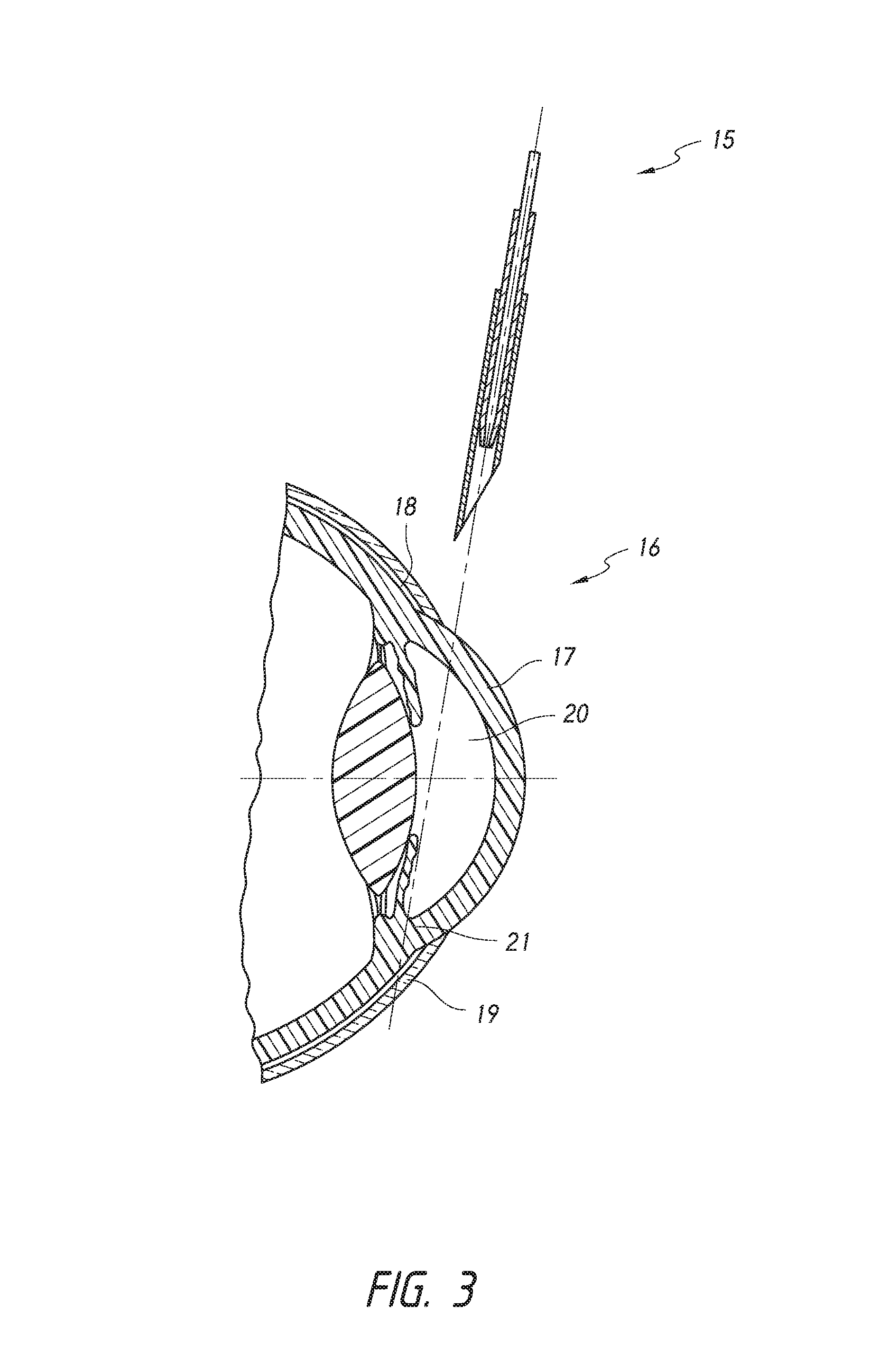
[0136]The present inventions generally relate to methods for treating closed angle glaucoma that involve using a deployment device that is configured to both re-open a partially or completely closed anterior chamber angle and deploy an intraocular shunt. In certain aspects, some methods involve inserting into an eye a deployment device configured to hold an intraocular shunt, using the device to re-open an at least partially closed anterior chamber angle of an eye, and deploying the shunt from the device.

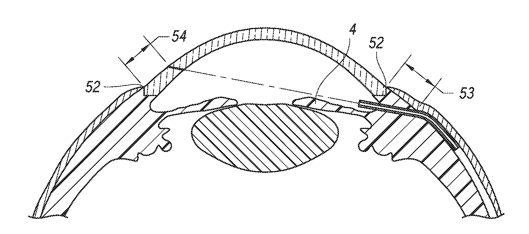
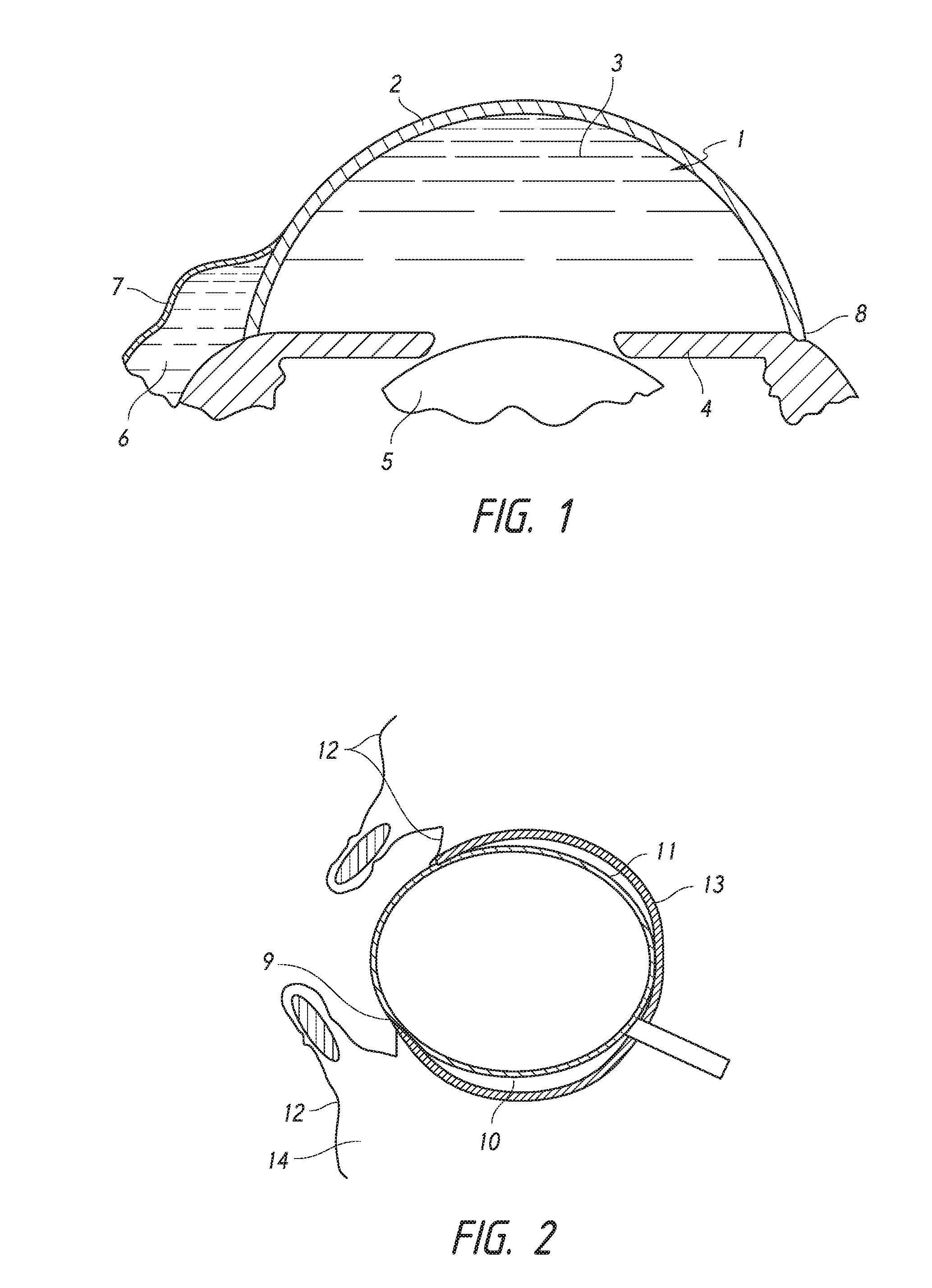
[0137]FIG. 1 provides a schematic diagram of the general anatomy of the eye. An anterior aspect of the anterior chamber 1 of the eye is the cornea 2, and a posterior aspect of the anterior chamber 1 of the eye is the iris 4. Beneath the iris 4 is the lens 5. The anterior chamber 1 is filled with aqueous humor 3. The aqueous humor 3 drains into a space(s) 6 below the conjunctiva 7 through the trabecular meshwork (not shown in detail) of the sclera 8. The aqueous humor is drained from...


PUM


Abstract

Glaucoma can be treated by implanting an intraocular shunt into the eye. Such procedures can employ various deployment devices, shunts, and implantation techniques. A method for treating glaucoma can include positioning an intraocular shunt in eye tissue, without use of an optical apparatus that contacts the eye, such that the shunt conducts fluid from the anterior chamber of the eye and delivers a pharmaceutical or biological agent to the eye.

Description

CROSS-REFERENCE TO RELATED APPLICATIONS[0001]This application is a continuation of U.S. patent application Ser. No. 14 / 313,970, filed Jun. 24, 2014; U.S. patent application Ser. No. 14 / 313,970 is a continuation-in-part of U.S. patent application Ser. No. 12 / 946,572, filed on Nov. 15, 2010, now U.S. Pat. No. 8,852,256; U.S. patent application Ser. No. 14 / 313,970 is also a continuation-in-part of U.S. patent application Ser. No. 12 / 946,222, filed on Nov. 15, 2010, now abandoned; U.S. patent application Ser. No. 14 / 313,970 is also a continuation-in-part of U.S. patent application Ser. No. 12 / 946,240, filed on Nov. 15, 2010, now U.S. Pat. No. 8,828,070; U.S. patent application Ser. No. 14 / 313,970 is also a continuation-in-part of U.S. patent application Ser. No. 12 / 946,251, filed on Nov. 15, 2010, now U.S. Pat. No. 9,095,411; U.S. patent application Ser. No. 14 / 313,970 is also a continuation-in-part of U.S. patent application Ser. No. 12 / 946,263, filed on Nov. 15, 2010, now U.S. Pat. No...

Claims


Application Information

Patent Timeline
Patent Type & Authority Applications(United States)
IPC IPC(8): A61F9/007A61B17/34
CPCA61B17/3415A61F9/00781A61B17/3468A61B17/3417A61F9/0008A61F9/0017A61F9/007A61M27/002
Inventor HORVATH, CHRISTOPHERROMODA, LASZLO O.GRABNER, GUENTHERREITSAMER, HERBERT A.
Owner AQUESYS INC
Why Eureka
Try Eureka