Registration of medical images

Active Publication Date: 2016-09-22
KONINKLJIJKE PHILIPS NV
View PDF7 Cites 6 Cited by
  • Summary
  • Abstract
  • Description
  • Claims
  • Application Information

AI Technical Summary

Benefits of technology

[0008]During a registration procedure, in which a 2D image such as an X-ray image is aligned/registered with a 3D image (or the 2D projection thereof), the X-ray image is filtered in a way that bony landmarks in the X-ray image may be boosted such that these landmarks become more visible in the blended/overlaid image. This may serve to better visualize to an operator, if the overlap is optimal, thereby helping the ope

Problems solved by technology

Low contrast features in X-ray images, such as the edges of spinal discs or the outlines of thoracic bones, may be difficult to distinguish

Method used


Image

  • Registration of medical images
  • Registration of medical images
  • Registration of medical images

Examples


Example

[0018]The reference symbols used in the drawings, and their meanings, are listed in summary form in the list of reference signs. In principle, identical parts are provided with the same reference symbols in the figures.

DETAILED DESCRIPTION OF EMBODIMENTS

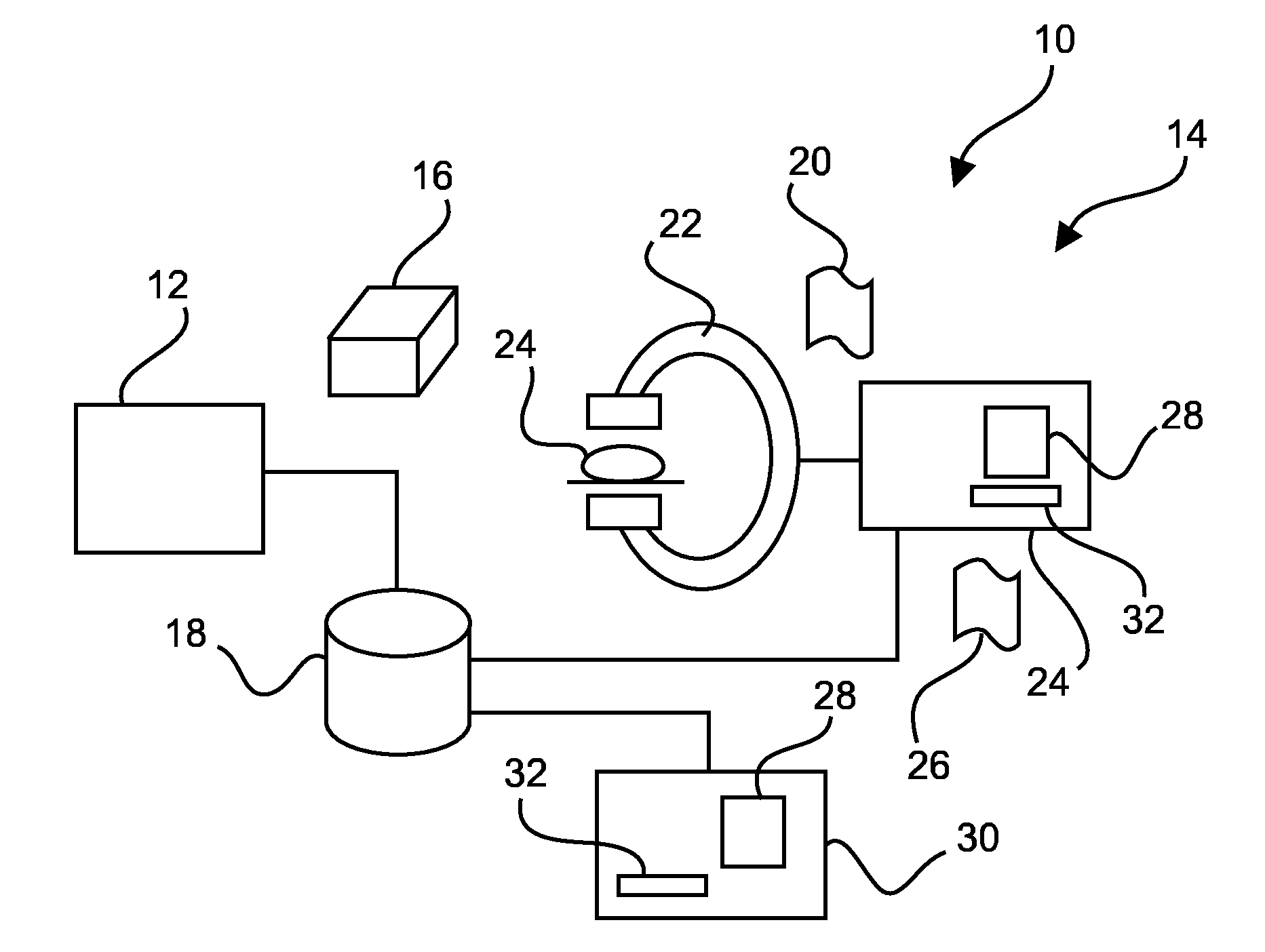
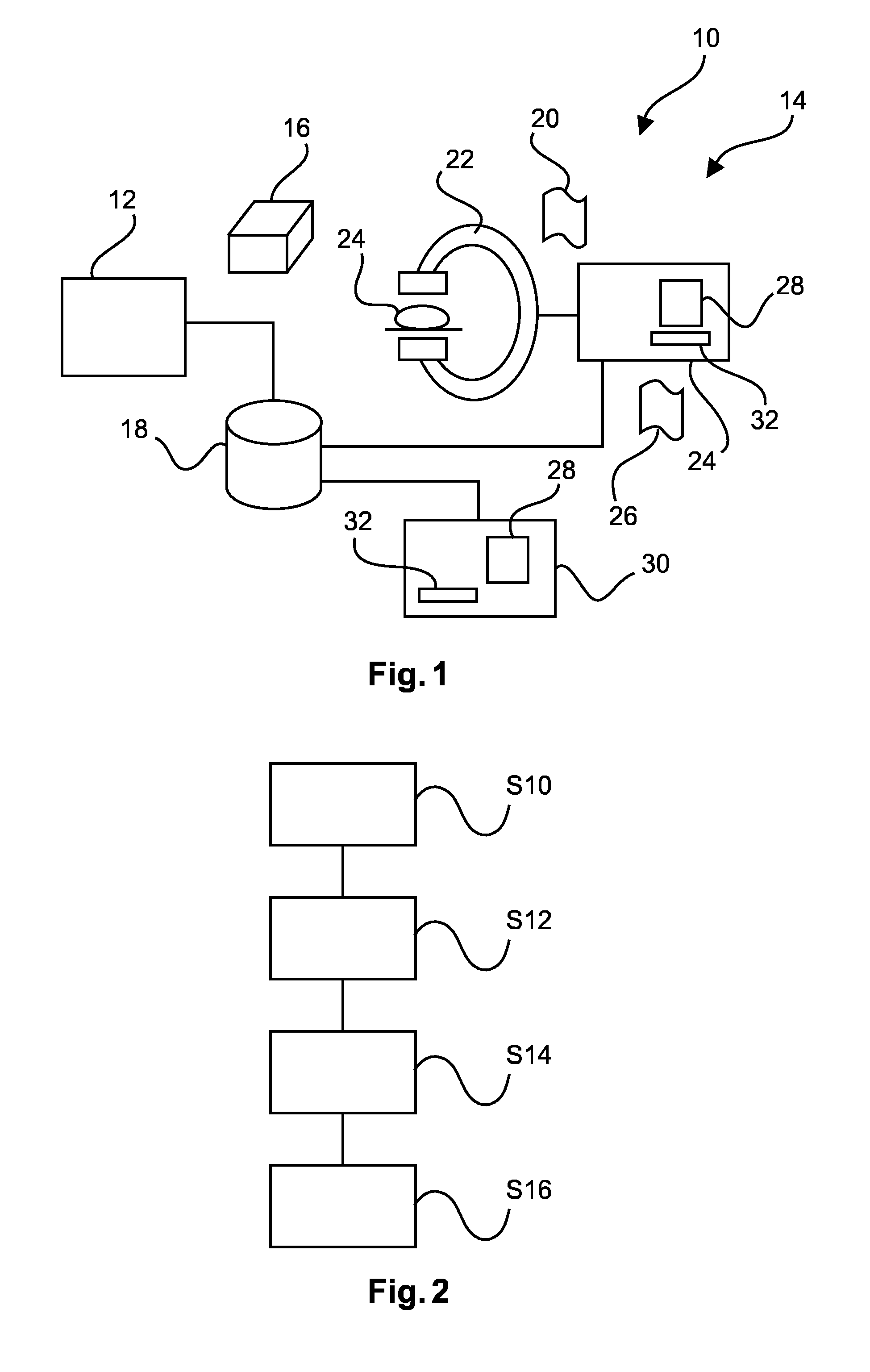
[0019]FIG. 1 schematically shows a system 10 comprising a 3D imaging device 12, and a 2D imaging device 14.

[0020]The 3D imaging device 10 may be a CT (computer tomography) or MRT (magnet resonance tomography) device and / or may be located remote from the 2D imaging device 12. For example, both devices may be situated in different rooms of a clinic or at different doctor's offices.

[0021]The 3D imaging devices is adapted for generating 3D images 16. A 3D image 16 may comprise voxels, each voxel comprising at least one intensity value associated to three coordinates. The 3D image data 16 may be acquired at a different time as the 2D imaging device 12 is used and / or may be stored in database 18.

[0022]The 2D imaging device 14 may be an X-r...


PUM


Abstract

A method for registration of medical images comprises: receiving a 2D X-ray image (20) acquired with a medical 2D imaging device (14) under a first view direction; filtering the 2D X-ray image (20), such that high frequency components of the 2D X-ray image are emphasized with respect to low frequency components of the 2D X-ray image; receiving a 3D image (16) acquired with a medical 3D imaging device (12); generating a 2D projection image (26) from the 3D image, wherein the 2D projection image is generated with a second view direction; overlaying the filtered 2D X-ray image and the 2D projection image; providing functionality for changing the second view direction, such that the 2D projection image is registered with the filtered 2D X-ray image.

Description

FIELD OF THE INVENTION[0001]The invention relates to a method, a computer program, a computer-readable medium and a device for registration medical images.BACKGROUND OF THE INVENTION[0002]During a procedure that uses multiple imaging modalities, for example CT and live X-ray, a physician may need to register the modalities. For example, when using an interventional X-ray device, in particular a C-arm device, the physician may move the C-arm to a certain angulation that gives him a clear view on landmarks in a body of a patient that can be used for registration, for example the pelvic bone. He will then acquire an exposure run or a single shot image. Next, the physician may move the C-arm to a different angulation that may be sufficiently different (e.g. more than 45 degrees difference) and shoot another exposure or single shot.[0003]Both X-ray runs may be shown in different viewports on a display device, where they are blended or overlaid with projection images of the CT volume (3D)...

Claims


Application Information

Patent Timeline
IPC IPC(8): G06T7/00
CPCG06T7/0028G06T7/0012G06T2207/10088G06T2207/10124G06T2200/04G06T2207/10081G06T2207/10072G06T7/33A61B6/5247G06T11/008G06V2201/12
Inventor ELENBAAS, THIJSDEN HARTOG, MARKUS JOHANNES HARMENVAN GELOOF, MARTIJNDE PAUS, ROBIN PIETER
Owner KONINKLJIJKE PHILIPS NV
Why Eureka
Try Eureka