X-ray ct apparatus and contrast imaging method

a technology of contrast imaging and ct apparatus, which is applied in the field of x-ray ct apparatus and contrast enhanced scanning method, can solve the problems of heavy patient burden and examination needs to be performed, and achieve the effect of satisfying the contrast

Inactive Publication Date: 2017-01-05
HITACHI LTD
View PDF6 Cites 15 Cited by
  • Summary
  • Abstract
  • Description
  • Claims
  • Application Information

AI Technical Summary

Benefits of technology

[0009]The present invention can provide an X-ray CT apparatus and a contrast enhanced scanning method that can obtain an image having a satisfactory contrast eff...

Problems solved by technology

Continuing scanning in the state where the scanning position has overtaken the contrast medium cannot obtain a desired contrast...

Method used


Image

  • X-ray ct apparatus and contrast imaging method
  • X-ray ct apparatus and contrast imaging method
  • X-ray ct apparatus and contrast imaging method

Examples


Embodiment Construction

[0018]Hereinafter, an embodiment of the present invention will be described in detail by referring to the drawings.

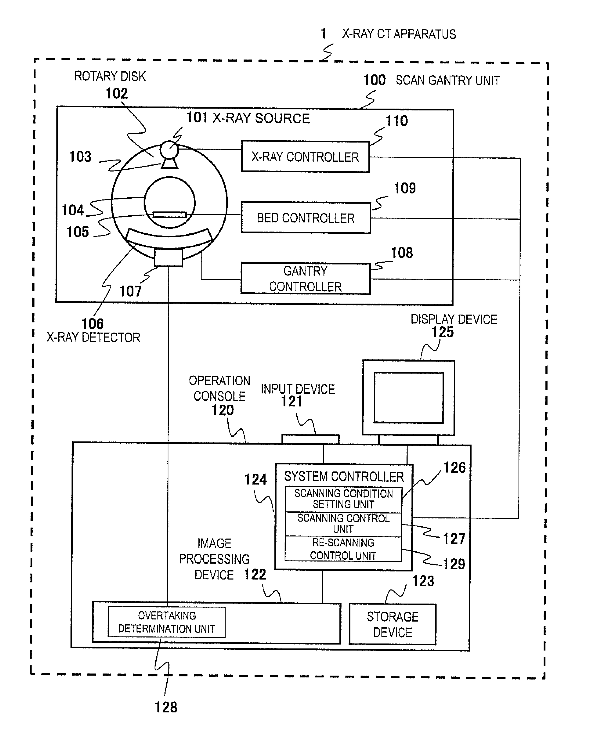
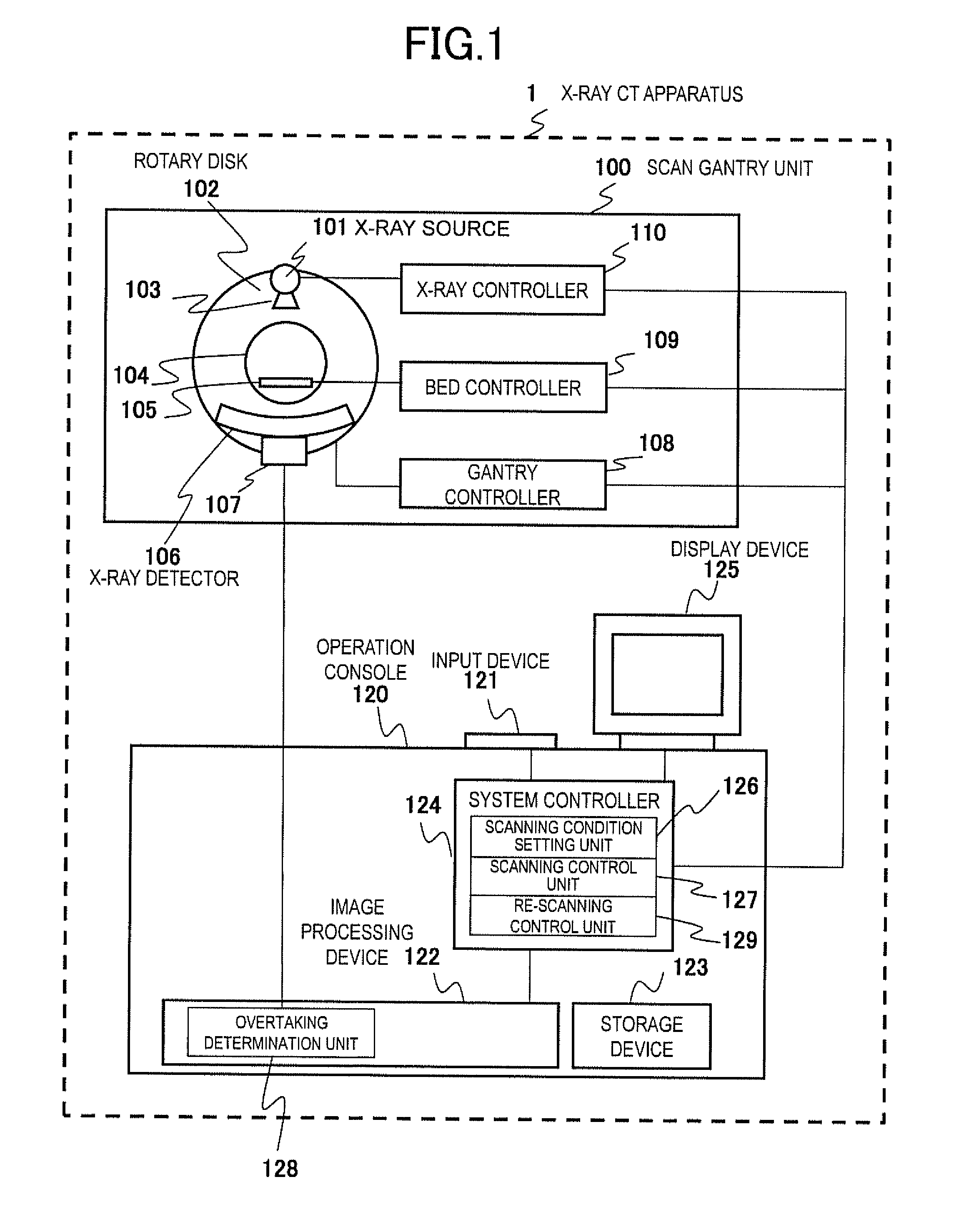
[0019]First, referring to FIG. 1, the overall configuration of the X-ray CT apparatus 1 will be described.

[0020]As shown in FIG. 1, the X-ray CT apparatus 1 comprises a scan gantry unit 100, a bed 105, and an operation console 120. The scan gantry unit 100 is a device that irradiates X-rays to an object and detects the X-rays transmitted through the object. The operation console 120 is a device that controls each part of the scan gantry unit 100 and acquires the transmitted X-ray data measured by the scan gantry unit 100 to generate an image. The bed 105 is a device that places the object and carries the object in and out of an X-ray irradiation range of the scan gantry unit 100.

[0021]The scan gantry unit 100 comprises an X-ray source 101, a rotary disk 102, a collimator 103, an X-ray detector 106, a data acquisition system 107, a gantry controller 108, a bed controller...


PUM


Abstract

In order to provide an X-ray CT apparatus and a contrast enhanced scanning method that can obtain an image having a favorable contrast effect without re-injecting contrast medium even when a scanning position overtakes a position of the contrast medium during scanning using the contrast medium, the X-ray CT apparatus determines whether or not a current scanning position overtakes the contrast medium during main scanning, resets a scanning condition when overtaking the contrast medium, executes re-scanning under the reset scanning condition, and determines overtaking of the contrast medium based on differential data between measurement data acquired by non-contrast enhanced scanning to be performed before the main scanning and measurement data to be acquired by the main scanning. Hence, no image needs to be reconstructed during scanning to check whether or not there is a contrast effect, and the overtaking determination is performed at a high speed, which can swiftly shift to re-scanning.

Description

TECHNICAL FIELD[0001]The present invention relates to an X-ray CT apparatus and a contrast enhanced scanning method and, in detail, to the X-ray CT apparatus that can perform scanning using contrast medium.BACKGROUND ART[0002]Conventionally, in an examination using an X-ray CT (Computed Tomography) apparatus, contrast CT examinations that perform scanning while contrast medium is being injected into an object are performed in order to obtain images with shades appropriate for diagnosis. In the contrast CT examinations, the injected contrast medium is delivered to the entire body through the blood flow, and when the contrast medium arrives at a scanning site, scanning using the X-ray CT apparatus starts. Whether or not the contrast medium has arrived at the scanning site is determined by, for example, a monitoring scan that monitors a density change of the contrast medium in a predetermined region of interest. Patent Literature 1 discloses a technique for measuring a density (CT valu...

Claims


Application Information

Patent Timeline
IPC IPC(8): A61B6/02A61B6/00A61B6/03
CPCA61B6/027A61B6/541A61B6/035A61B6/032A61B6/481A61B6/54
Inventor GOTO, TAIGA
Owner HITACHI LTD
Why Eureka
Try Eureka