Toroidal glaucoma drainage device

a glaucoma and drainage device technology, applied in the field of implantable devices for ocular drainage, can solve the problems of hardly tolerable side effects, failure to control eye pressure, late-onset blebs,

Inactive Publication Date: 2017-03-16
TEL HASHOMER MEDICAL RES INFRASTRUCTURE & SERVICES
View PDF7 Cites 13 Cited by
  • Summary
  • Abstract
  • Description
  • Claims
  • Application Information

AI Technical Summary

Benefits of technology

[0014]The invention is directed to a glaucoma drainage device for intraocular implanting, comprising a tubing having an outer surface impermeable to aqueous fluids with a plurality of laser-sensitive spots permeable to aqueous fluids after laser irradiation, wherein free ends of the tubing are attached to each other by means of a lock to form a closed loop, the device being implanted in the anterior chamber near the limbus of an eye and being sutured to the sclera to ensure its fixed position within the eye, said lock optionally comprising a T-tube to connect said loop with an extraocularly situated plate via an essentially linear connector, wherein a part of said loop is located within the anterior chamber of said eye shunting fluid out of the anterior chamber, the volume of said fluid being affected by the fraction of said spots being converted to openings by said irradiation, the fraction being optimized by sequentially irradiating the device in several increments. The glaucoma drainage device according to the invention is mainly used in managing refractory glaucoma.
[0015]The invention provides a method of treating refractory glaucoma in an eye in need of such treatment, comprising i) providing an intraocular tubular implant having an outer surface impermeable to aqueous fluids with a plurality of laser-sensitive spots permeable to aqueous fluids after laser irradiation, the implant having a shape of open tubing optionally having a narrowed site (a neck); ii) implanting said tubular implant in the anterior chamber, extraocularly near the limbus or intraocularly at the level of anterior chamber angle; iii) attaching the free ends of said tubular implant to each other by means of a lock to form a closed loop, the lock optionally having a T-shape, the loop essentially having a toroidal shape; iv) fixing the position of said loop inside the eye, but under the conjunctiva or scleral flap, by suturing it to the sclera, optionally using said narrowed site (neck), wherein the loop is near the limbus of an eye, its part being located within the anterior chamber; v) if the loop comprises said T-shaped lock, connecting it with an extraocularly situated plate via a linear connector, the plate being sutured to the sclera, located outside the sclera / below scleral patch of donor tissue; vi) measuring the intraocular pressure (TOP) of said eye; vii) if said TOP is higher than required, irradiating the implanted device by laser whereby opening a part of said plurality of laser-sensitive spots on the surface of the loop; and viii) examining whether the step vii resulted in lowering the value of said TOP; ix) repeating steps vii and viii till the TOP value reached a desired value.

Problems solved by technology

Eye drops or oral medications are employed to lower the eye pressure, but they often do not succeed in controlling eye pressure or they result in hardly tolerable side effects, necessitating surgery.
Many of the serious side effects or complications that occur with the procedure involve blister formation, blebs, which cause leakage and may eventually cause blindness.
Some drugs used to prevent post-operational scarring unfortunately cause late-onset leaking blebs, requiring lifelong monitoring after surgery.
Another complication in up to one third of patients is developing adhesive-like substances which may glue the iris to the cornea.
Complications after such implantation may include cataract, detached retina, breakdown of the cornea, bleeding, and hypotony, the latter occurring when the outflow is excessive.
Other risks and problems include eye movement disorders, such as double vision and strabismus, or implant blockage, requiring additional surgery.
Some techniques are too risky and are reserved only for people with end-stage glaucoma.
Many techniques require extraordinary surgical skill, and even if performed perfectly, they do not always sufficiently handle the IOPs.
Special problems, like for example combined cataract with glaucoma, require special attitudes, and despite their variability, the available techniques frequently cannot provide an efficient solution.
The free inner end has a great potential to multiple complications—it can move, be displaced and eventually touch surfaces of the cornea and iris resulting in many problems, like corneal endothelial cells loss, pigment dispersion syndrome, inflammation, hemorrhages, failure, cataracts, etc.
Undesired phenomena include, on one hand, blocking of the drainage flow, and, on the other hand, excessive drainage; the liquid outflow can be adjusted to the observed clinical IOP only with great difficulties.
Among other drawbacks, said available devices are disadvantageously exposed on the cornea surface, and moreover have very limited and mostly unpredictable means to control the eye fluid flows.

Method used


Image

  • Toroidal glaucoma drainage device
  • Toroidal glaucoma drainage device
  • Toroidal glaucoma drainage device

Examples


Embodiment Construction

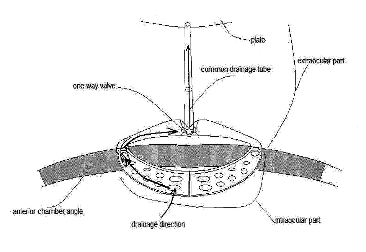
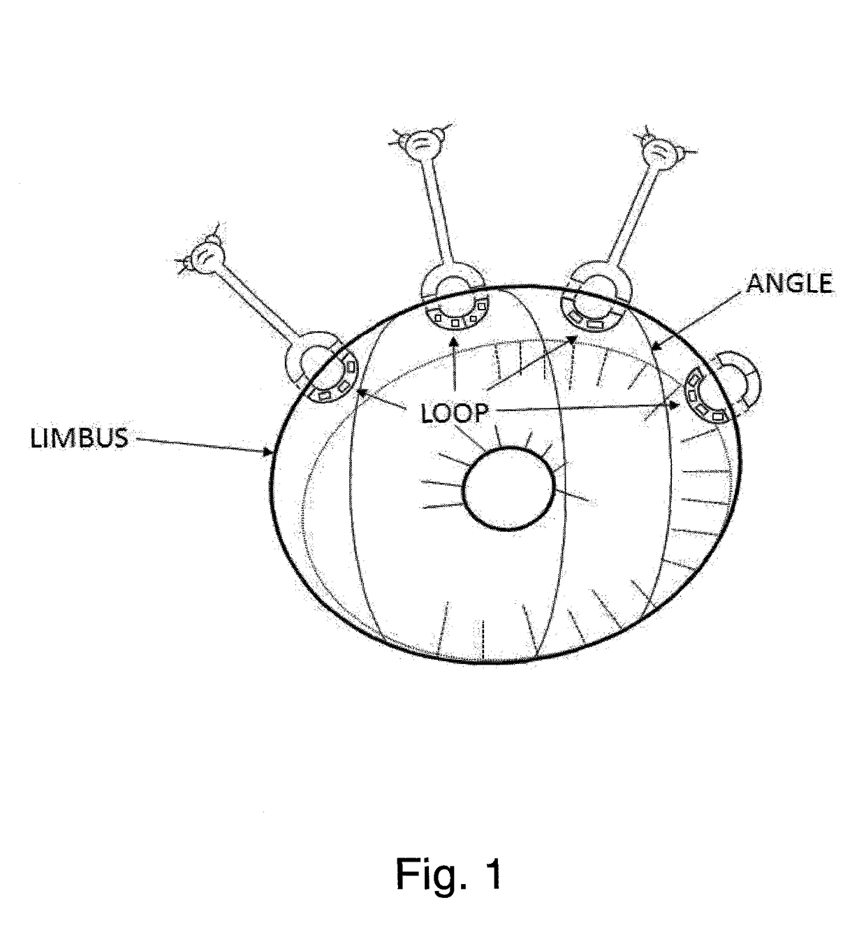
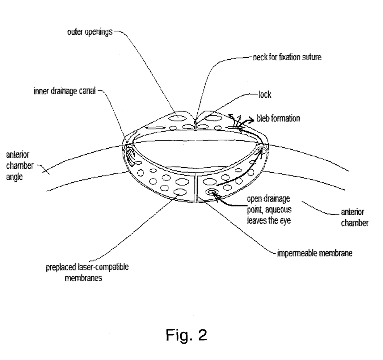
[0022]An intraocular drainage device has now been designed that enables to adjust the amount of the exiting liquid according to observed intraocular pressure values, so that glaucoma problems may be more easily managed than with known devices. The device has a torus-shaped outer surface comprising an impermeable and laser insensitive membrane on which a plurality of laser-sensitive spots are placed, potentially forming a plurality of exits for the eye fluid after being eventually opened by applying YAG laser light—according to the need. The laser-sensitive spots are preformed during manufacturing of the device and exist before implanting the device to the eye. Preferably, one or more devices are placed in the angle area of the eye suffering from glaucoma, near / through the limbus. More spots, preformed on the torus surface, are laser-irradiated, resulting in higher permeability of the toroidal device or devices, providing higher fluid flow. This enables fine regulation of the drainag...


PUM


Abstract

The invention provides an intraocular drainage implant whose drainage capacity is regulated by opening a plurality of laser-sensitive spots. The implant is useful for fine regulation of the eye fluid outflow in handling glaucoma.

Description

FIELD OF THE INVENTION[0001]The present invention relates to an implantable device for ocular drainage useful in handling glaucoma. The device and methods of use thereof enable fine regulation of the eye fluid outflow.BACKGROUND OF THE INVENTION[0002]Glaucoma is a widespread eye disease, afflicting more than two million people in the U.S. alone and causing blindness in 120,000 of them. The only modifiable risk factor of glaucoma is high intraocular pressure (IOP), thus all the treatment options try to control it. Eye drops or oral medications are employed to lower the eye pressure, but they often do not succeed in controlling eye pressure or they result in hardly tolerable side effects, necessitating surgery. The surgical procedures usually include trabeculectomy aiming at opening the full thickness of the drainage area, or laser trabeculoplasty that partially opens the drainage area. Trabeculectomy, with small modifications, has been used for hundred of years. Many of the serious s...

Claims


Application Information

Patent Timeline
Patent Type & Authority Applications(United States)
IPC IPC(8): A61F9/007A61F9/008
CPCA61F9/008A61F9/00781A61F2/15A61F9/00821A61F2230/0063
Inventor PIVEN, ILIA
Owner TEL HASHOMER MEDICAL RES INFRASTRUCTURE & SERVICES
Why Eureka
Try Eureka