
Quantitative tissue property mapping for real time tumor detection and interventional guidance

a tissue property and real-time technology, applied in the field of medical imaging, can solve the problems of large positional errors of patient's movements, lack of real-time continuous guidance, time-consuming, etc., and achieve the effects of reducing the influence of depth-dependent effects, avoiding potential injury, and high computational efficiency and accuracy

Pending Publication Date: 2017-03-30
THE JOHN HOPKINS UNIV SCHOOL OF MEDICINE
View PDF3 Cites 5 Cited by
  • Summary
  • Abstract
  • Description
  • Claims
  • Application Information

AI Technical Summary

Benefits of technology

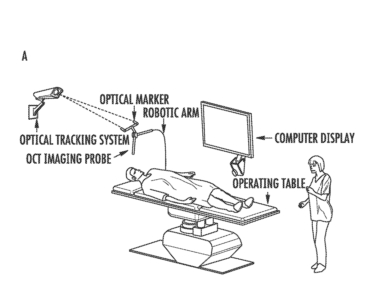
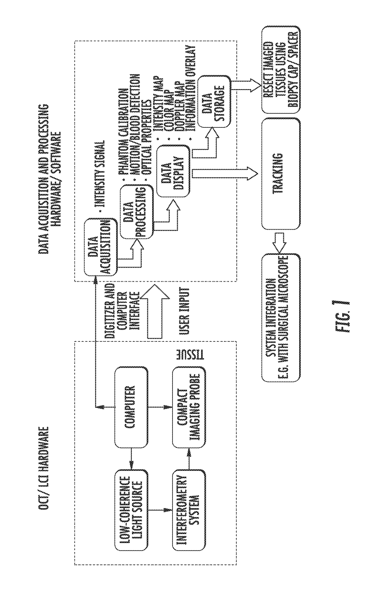
The present invention is a method and system for analyzing optical properties of tissues and tumors in real-time using high-resolution imaging techniques. The method includes programming the steps on non-transitory computer-readable media, and utilizing exponential and Frequency-domain fitting methods for accurate and reliable characterization of optical properties with high computational efficiency and accuracy. The method also mitigates the influence of depth-dependent effects of the beam profile by creating phantoms and calibrating the imaging data. The system includes an optical imaging device integrated with a tracking system for real-time position and orientation tracking, as well as an aiming beam for interventional guidance. The method also includes the use of graphics processing unit (GPU)-based or field-programmable gate array (FPGA)-based parallel processing algorithms for optimal computational efficiency and real-time acquisition, processing, and displays of tissue optical properties, structures, and blood flow. The technical effects of this invention include improved identification and minimization of cancer tissues and tracking of tumor borders during surgery, facilitating tumor resection and providing accurate imaging-histological correlations for basic science and clinical research purposes.

Problems solved by technology

For example, surgical navigations based on pre-operative MRI is the current standard of care for brain cancer, but causes large positional errors from the patient's motions e.g. breathing and heartbeat.
Intra-operative MRI provides better resolution and accuracy, but does not provide real-time continuous guidance; it is also time consuming and often costs millions of dollars per unit, which only few hospitals can afford.
Ultrasound is portable and low-cost, but its use in the operating room is limited for certain cancer applications due to insufficient tissue contrast and resolution.

Method used


Image

  • Quantitative tissue property mapping for real time tumor detection and interventional guidance
  • Quantitative tissue property mapping for real time tumor detection and interventional guidance
  • Quantitative tissue property mapping for real time tumor detection and interventional guidance

Examples


Embodiment Construction

[0020]The presently disclosed subject matter now will be described more fully hereinafter with reference to the accompanying Drawings, in which some, but not all embodiments of the inventions are shown. Like numbers refer to like elements throughout. The presently disclosed subject matter may be embodied in many different forms and should not be construed as limited to the embodiments set forth herein; rather, these embodiments are provided so that this disclosure will satisfy applicable legal requirements. Indeed, many modifications and other embodiments of the presently disclosed subject matter set forth herein will come to mind to one skilled in the art to which the presently disclosed subject matter pertains having the benefit of the teachings presented in the foregoing descriptions and the associated Drawings. Therefore, it is to be understood that the presently disclosed subject matter is not to be limited to the specific embodiments disclosed and that modifications and other ...


PUM


Abstract

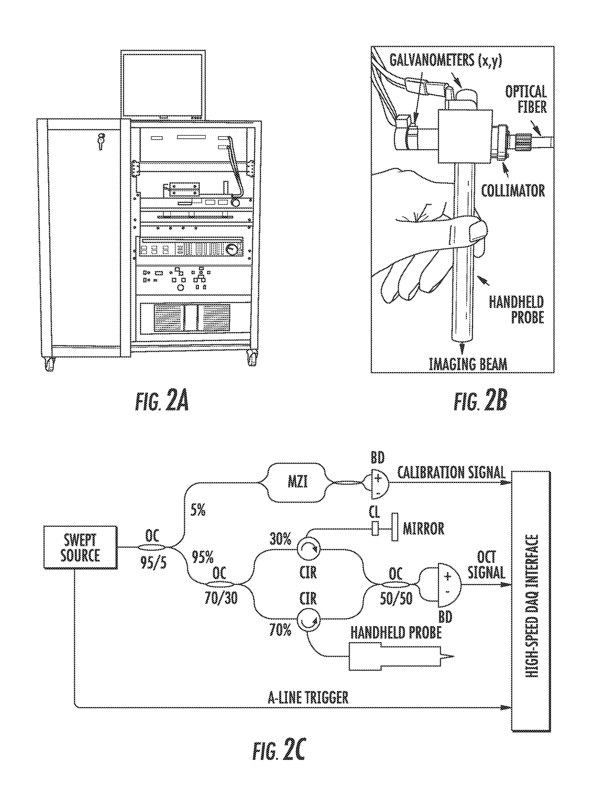
The present invention is directed to a method for real-time characterization of spatially-resolved tissue optical properties using OCT / LCI. Imaging data are acquired, processed, displayed and stored in real-time. The resultant tissue optical properties are then used to determine the diagnostic threshold and to determine the OCT / LCI detection sensitivity and specificity. Color-coded optical property maps are constructed to provide direct visual cues for surgeons to differentiate tumor versus non-tumor tissue. These optical property maps can be overlaid with the structural imaging data and / or Doppler results for efficient data display. Finally, the imaging system can also be integrated with existing systems such as tracking and surgical microscopes. An aiming beam is generally provided for interventional guidance. For intraoperative use, a cap / spacer may also be provided to maintain the working distance of the probe, and also to provide biopsy capabilities. The method is usable for research and clinical diagnosis and / or interventional guidance.

Description

CROSS REFERENCE TO RELATED APPLICATIONS[0001]This application claims the benefit of U.S. Provisional Patent Application No. 61 / 970,104 filed Mar. 25, 2014, which is incorporated by reference herein, in its entirety.GOVERNMENT RIGHTS[0002]This invention was made with government support under R01EB007636, R01CA120480, and R01NS070024 awarded by the National Institutes of Health. The government has certain rights in the invention.FIELD OF THE INVENTION[0003]The present invention relates generally to medical imaging. More particularly, the present invention relates to a method for Optical Coherence Tomography (OCT) or low coherence interferometry (LCI) imaging based tumor detection and interventional guidance.BACKGROUND OF THE INVENTION[0004]Approximately 1,665,540 new cancer cases and 585,720 cancer deaths occur annually in the United States. Surgery is the primary method of treatment for most isolated solid cancers and often plays a role the prolongation of survival. Previous studies ...

Claims


Application Information

Patent Timeline
IPC IPC(8): A61B5/00A61B90/20
CPCA61B5/0073A61B5/00A61B90/20A61B5/0086A61B5/7425A61B5/0066A61B2090/3735
Inventor LI, XINGDEQUINONES-HINOJOSA, ALFREDOMCVEIGH, ELLIOTCHAICHANA, KAISORNKUT, CARMENXI, JIEFENG
Owner THE JOHN HOPKINS UNIV SCHOOL OF MEDICINE
Learn More