Visualization, navigation, and planning with electromagnetic navigation bronchoscopy and cone beam computed tomography integrated

a technology of electromagnetic navigation and computed tomography, which is applied in the field of assisting surgeons, can solve the problems of not providing guidance, one or more portions of a patient's lungs may lose their ability to function normally, and system does not account for changes that may have occurred after the ct scan, or for movement during the treatment procedur

Active Publication Date: 2017-06-08
TYCO HEALTHCARE GRP LP
View PDF8 Cites 12 Cited by
  • Summary
  • Abstract
  • Description
  • Claims
  • Application Information

AI Technical Summary

Benefits of technology

[0015]In a further aspect of the present disclosure, the EM field generator compensa

Problems solved by technology

Pulmonary disease may cause one or more portions of a patient's lungs may lose its ability to function normally and thus may need to be treated.
Traditional pre-operative images are helpful, to an extent, with the for

Method used


Image

  • Visualization, navigation, and planning with electromagnetic navigation bronchoscopy and cone beam computed tomography integrated
  • Visualization, navigation, and planning with electromagnetic navigation bronchoscopy and cone beam computed tomography integrated
  • Visualization, navigation, and planning with electromagnetic navigation bronchoscopy and cone beam computed tomography integrated

Examples


Embodiment Construction

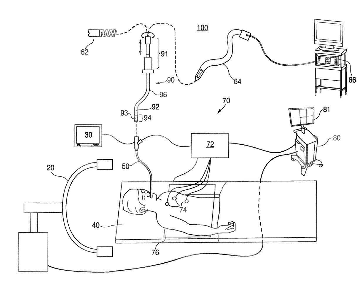
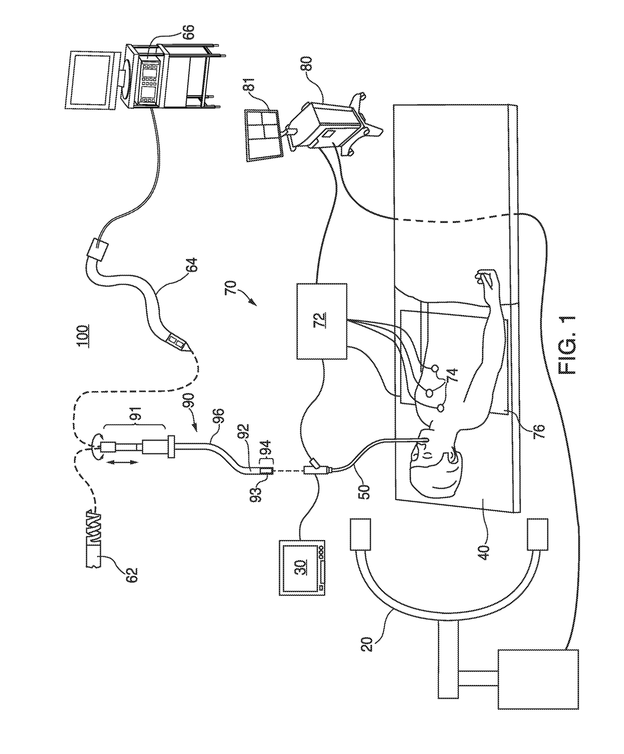
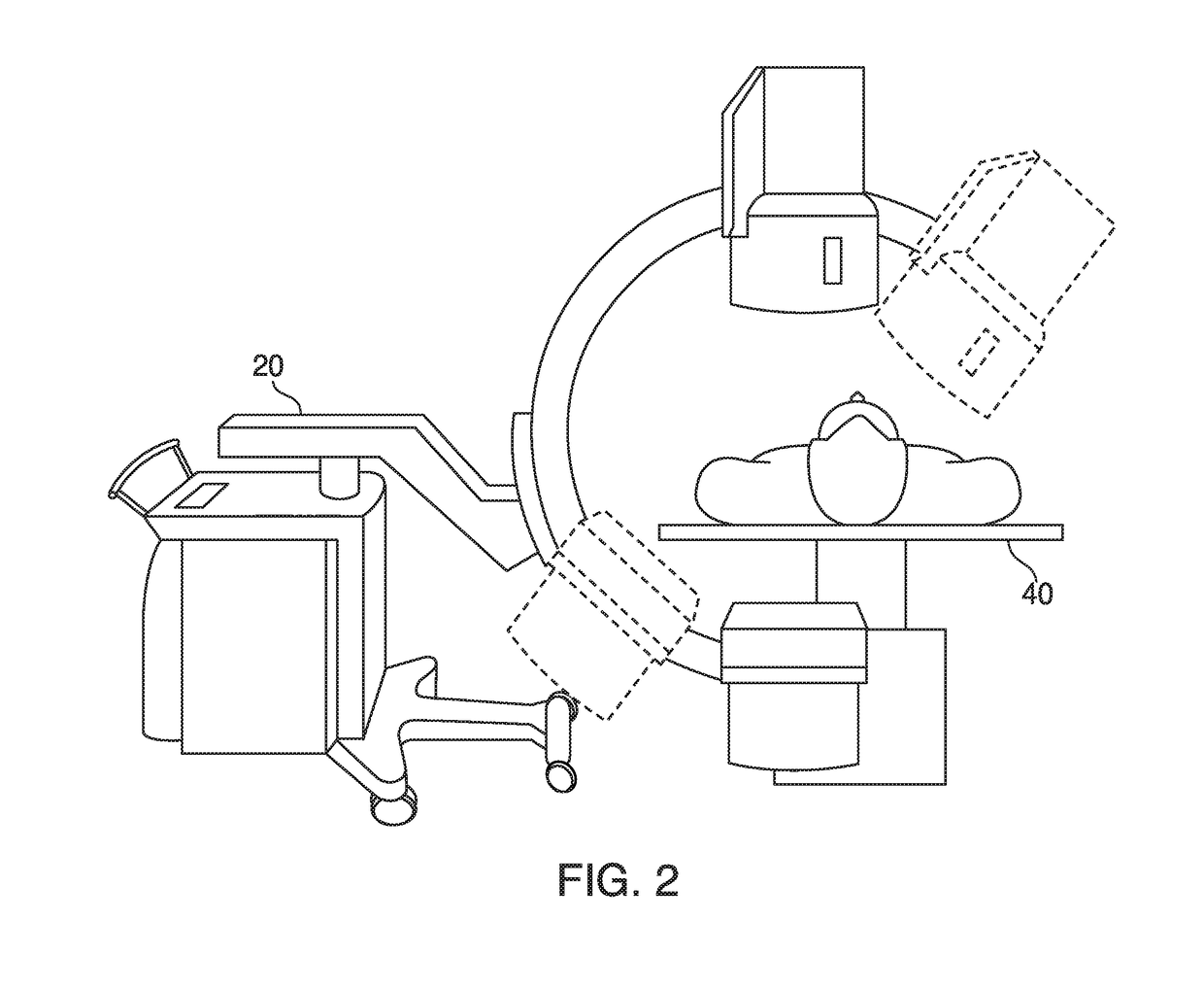
[0035]The present disclosure is directed to devices, systems, and methods for using cone beam computed tomography (CBCT) images while navigating tools in a patient's lungs and performing treatment in the patient's lungs. More particularly, the disclosure relates to integrating CBCT image data with a lung navigation system to update and / or improve localization and visualization of treatment targets and tools within the patient's lungs, and to update image-based guidance for a treatment procedure. The CBCT image data may be displayed in conjunction with or alongside a digital reconstruction, such as a three-dimensional (3D) model or map, of the patient's lungs, as well as image data from other imaging modalities including computed tomography (CT) images, magnetic resonance (MR) images, positron emission tomography (PET) images, fluoroscopic and other X-ray type images, and / or ultrasound images. The 3D model may be constructed based on preoperative image data from one or more of the af...


PUM


Abstract

The present disclosure relates to systems, devices and methods for providing visual guidance for navigating inside a patient's chest. An exemplary method includes presenting a three-dimensional (3D) model of a luminal network, generating, by an electromagnetic (EM) field generator, an EM field about the patient's chest, detecting a location of an EM sensor within the EM field, determining a position of a tool within the patient's chest based on the detected location of the EM sensor, displaying an indication of the position of the tool on the 3D model, receiving cone beam computed tomography (CBCT) image data of the patient's chest, detecting the location of the tool within the patient's chest based on the CBCT image data, and updating the indication of the position of the tool on the 3D model based on the detected location.

Description

CROSS-REFERENCE TO RELATED APPLICATIONS[0001]The present application claims the benefit of and priority to U.S. Provisional Patent Application Ser. No. 62 / 264,145 entitled VISUALIZATION, NAVIGATION, AND PLANNING WITH ELECTROMAGNETIC NAVIGATION BRONCHOSCOPY AND CONE BEAM COMPUTED TOMOGRAPHY INTEGRATED, filed on Dec. 7, 2015, by Haley et al., the entire contents of which is incorporated by reference herein.BACKGROUND[0002]Technical Field[0003]The present disclosure relates to tools for assisting surgeons during performance of medical procedures, and more particularly, to systems, devices, and methods for providing image-based guidance during medical procedures.[0004]Description of Related Art[0005]Pulmonary disease may cause one or more portions of a patient's lungs may lose its ability to function normally and thus may need to be treated. Lung treatment procedures may be very complex and would be greatly aided if the surgeon performing the procedure can visualize the way airways and ...

Claims


Application Information

Patent Timeline
IPC IPC(8): A61B6/12A61B6/00A61B18/12A61B10/02A61B18/14A61B18/18A61B6/03A61B5/06
CPCA61B6/12A61B6/032A61B6/4085A61B5/062A61B6/463A61B6/4441A61B2018/00541A61B18/1492A61B18/1815A61B18/1206A61B2018/1823A61B2018/00577A61B2018/00982A61B10/02A61B6/466A61B90/37A61B2090/3764A61B34/20A61B2034/2051A61B6/487A61B1/267G16H50/50A61B2017/00699
Inventor DICKHANS, WILLIAM J.HALEY, KAYLEN J.
Owner TYCO HEALTHCARE GRP LP
Why Eureka
Try Eureka