Apparatus for determining and quantifying the staining of ocular structures and method therefor

a technology of ocular structure and apparatus, applied in the field of surgical ophthalmology, can solve the problems of irreparably damaged retina, unpredictable process of manual introduction of dye inability to accurately predict the process of local resorption and local resorption by the capsule, etc., and achieve the effect of high contrast and suitable illuminating method

Inactive Publication Date: 2017-07-06
CARL ZEISS MEDITEC AG
View PDF6 Cites 4 Cited by
  • Summary
  • Abstract
  • Description
  • Claims
  • Application Information

AI Technical Summary

Problems solved by technology

If the desired layer of dye has gaps or at least relatively weakly stained zones, then firstly the desired treatment effect is not achieved as a result of a lack of absorption and, secondly, the laser radiation impinges on the retina with virtually no attenuation and can irreparably damage the retina.
If the desired dye layer contains gaps or at least less intensely dyed zones, then, due to the lacking absorption coefficient on the one hand, the desired treatment effect is not achieved and, on the other hand, the laser radiation hits the retina in a nearly undamped manner and can thus irreparably damage the retina.
What is disadvantageous in the current state of the art is that manual introduction of the dye and local resorption by the capsule are not comprehensively predictable processes.
In this regard, it may be the case that in the desired treatment zone only an insufficient amount of dye is applied or the latter is distributed non-uniformly.
Under certain circumstances, the surgeon might even totally fail to inject the dye.

Method used


Image

  • Apparatus for determining and quantifying the staining of ocular structures and method therefor
  • Apparatus for determining and quantifying the staining of ocular structures and method therefor
  • Apparatus for determining and quantifying the staining of ocular structures and method therefor

Examples


Embodiment Construction

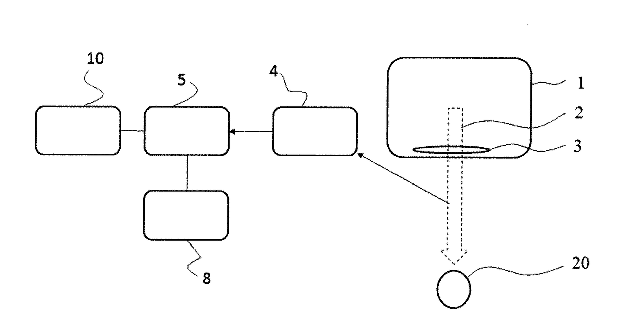
[0029]FIG. 1 shows a schematic illustration of a surgical microscope 1 with an apparatus for detecting and quantifying the staining of ocular structures according to the invention. The surgical microscope 1 includes a magnification unit 3, shown schematically. A line of sight 3 of a user through the surgical microscope 1 to a patient's eye 20 is depicted by the light path 2. The apparatus further includes an image capture sensor 4 for capturing images of an ocular structure and an evaluation module 5 configured to evaluate images taken by the image capture sensor 4 in accordance with a method of the invention.

[0030]In a first embodiment, the apparatus has an image capture sensor 4 which captures a reference image of a retro-illuminated unstained capsule. The reference image is preferably captured directly before staining in order to minimize any changes which may occur after the taking the reference image and prior to staining. After the ocular structure has been stained with a dye ...


PUM


Abstract

An apparatus has an image capture sensor which captures a reference image of a retro-illuminated unstained capsule of an eye. After the ocular structure has been stained with a dye for an ophthalmic surgical procedure a second image is captured as a measurement image via the image capture sensor. An evaluation module compares the reference image and the measurement image taken after the staining of the ocular structure. The evaluation module from the comparison of the reference and measurement images then determines the local light attenuation by the introduced dye. The staining of the eye can also be determined by only imaging a stained ocular structure of an eye and evaluating the images on the basis of predetermined acceptance values.

Description

FIELD OF THE INVENTION[0001]This invention relates to the field of surgical ophthalmology, especially cataract surgery using colorants and laser methods on the capsular bag and on the natural eye lens. The invention further relates to the field of refractive corneal surgery for the application and control of corneal tattoos.BACKGROUND OF THE INVENTION[0002]The application of dyes for staining and visualizing the anterior capsular surface in preparation for capsulorhexis has been established for years in cataract surgery. One of the most commonly used dyes for this clinical application is trypan blue. Trypan blue is very effective at dyeing the capsule as a result of its chemical binding characteristics and leads to a good visibility of the dyed structures due to its high absorption in the yellow to red spectral range.[0003]Capsulorhexis is an example for an eye operation in the anterior of an eye. Here, a piece of the anterior capsular bag of an eye is scored in a circular region an...

Claims


Application Information

Patent Timeline
Patent Type & Authority Applications(United States)
IPC IPC(8): A61B3/14A61B3/13A61B3/12A61B3/00
CPCA61B3/14A61B3/0025A61B3/12A61B3/13A61B3/0033A61B3/10G06T2207/30041G06T7/0014
Inventor GERLACH, MARIO
Owner CARL ZEISS MEDITEC AG
Why Eureka
Try Eureka