Combination of Anti-cs1 and Anti-pd1 antibodies to treat cancer (myeloma)

an antics1 and antipd1 antibody technology, applied in the field of myeloma, can solve the problems of relapse of almost all patients with multiple myeloma, debilitating side effects, fatigue, etc., and achieve the effects of synergistic treatment effect, good partial response, and stable diseas

Inactive Publication Date: 2017-12-14
BRISTOL MYERS SQUIBB CO
View PDF5 Cites 1 Cited by
  • Summary
  • Abstract
  • Description
  • Claims
  • Application Information

AI Technical Summary

Benefits of technology

[0068]The efficacy of the treatment methods provided herein can be assessed using any suitable means. In one embodiment, the treatment produces at least one therapeutic effect selected from the group consisting of complete response, very good partial response, partial response, and stable disease. In another embodiment, administration of an anti-PD1 antibody and an anti-CS1 antibody has a synergistic effect on treatment compared to administration of either antibody alone.

Problems solved by technology

However, despite these advances, almost all multiple myeloma patients relapse.
The increased production of M-protein has been linked to hyperviscosity syndrome in multiple myelomas, causing debilitating side effects, including fatigue, headaches, shortness of breath, mental confusion, chest pain, kidney damage and failure, vision problems and Raynaud's phenomenon (poor blood circulation, particularly fingers, toes, nose and ears).
These particles can block small blood vessels and cause pain and numbness in the toes, fingers, and other extremities during cold weather.

Method used


Image

  • Combination of Anti-cs1 and Anti-pd1 antibodies to treat cancer (myeloma)
  • Combination of Anti-cs1 and Anti-pd1 antibodies to treat cancer (myeloma)
  • Combination of Anti-cs1 and Anti-pd1 antibodies to treat cancer (myeloma)

Examples


example 1

Method for Cloning SLAMF7 cDNA into pFB Retroviral Vector

[0243]cDNA sequence from human SLAM family member 7 (hSLAMF7; synonyms: CS1-L) was cloned into retroviral vector encoding green fluorescent protein (GFP) (pFB-IRES-GFP, Stratagene).

[0244]The vector contains the murine leukemia retrovirus (MLV) packaging sequence and a multiple cloning site (MCS), flanked by the MLV long terminal repeat (LTR) regions.

[0245]The 5′ LTR functions as a strong promoter upon chromosomal integration of DNA. The pFB plasmid contains a cassette comprising an ECMV internal ribosome entry site (IRES) followed by a gene encoding GFP.

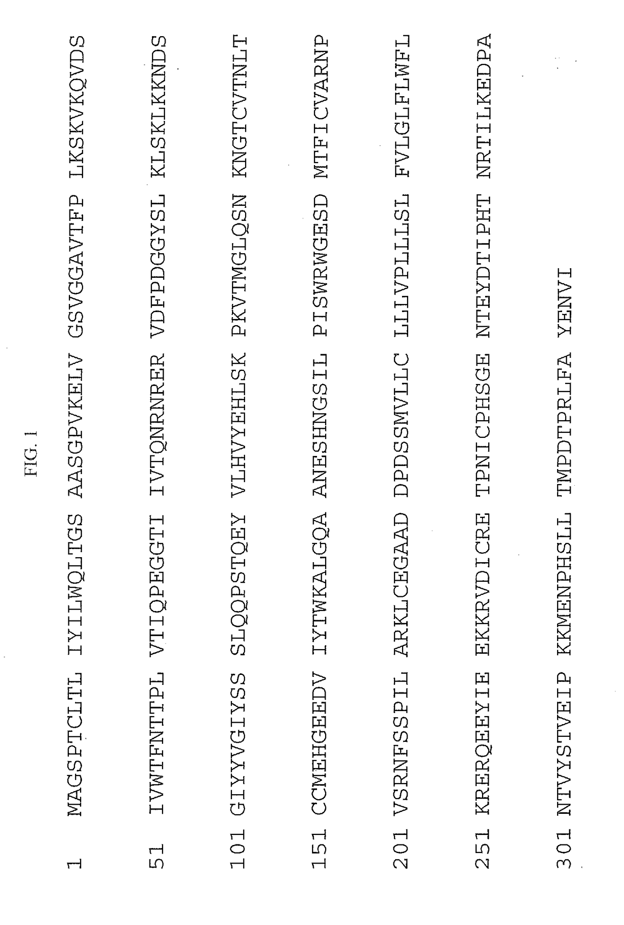
[0246]The cloned sequence of the encoded SLAMF7 protein sequence is provided in FIG. 1 (SEQ ID NO:7).

example 2

Method for Generation of A20 Mouse Tumor Cell Line Expressing Human SLAMF7

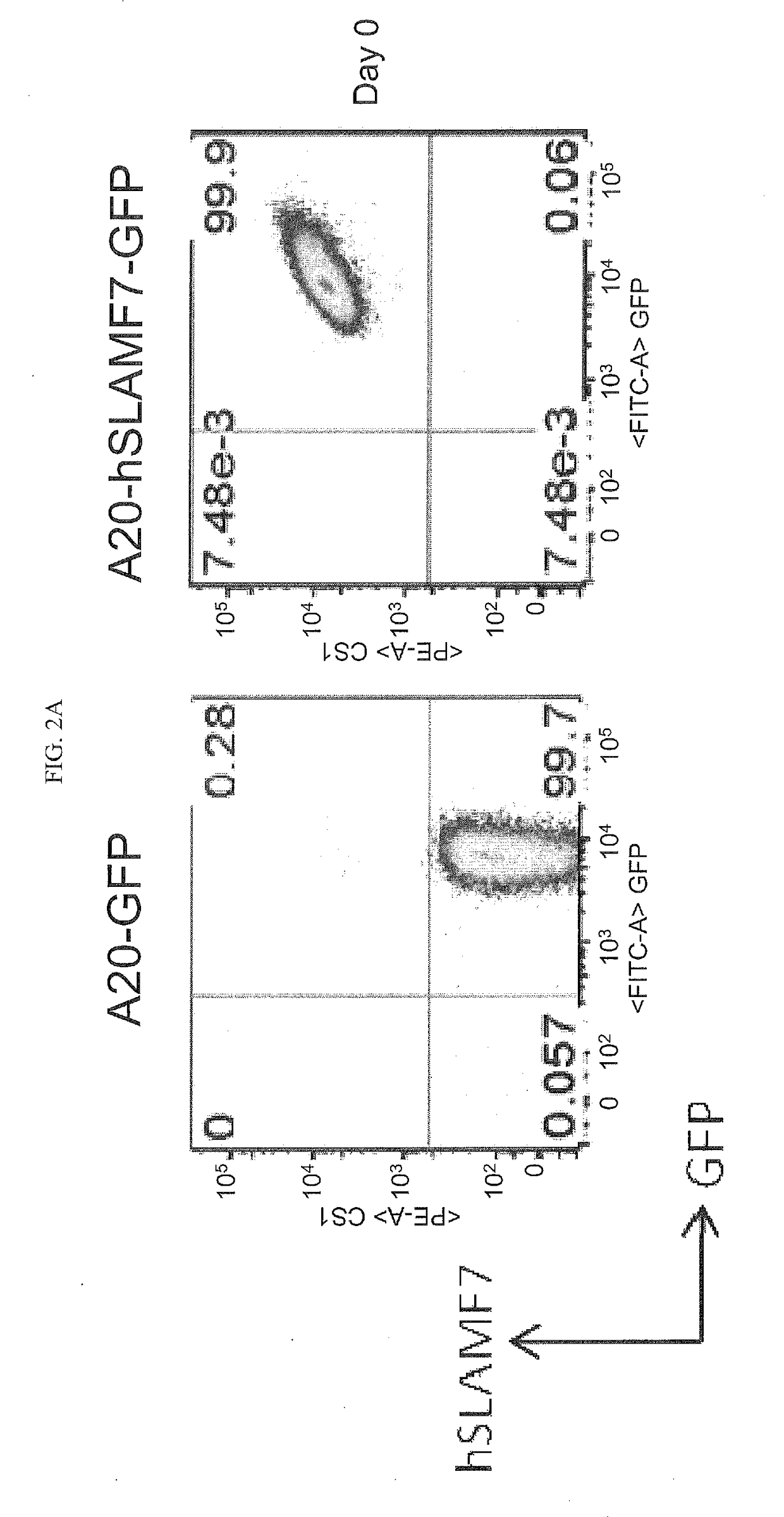
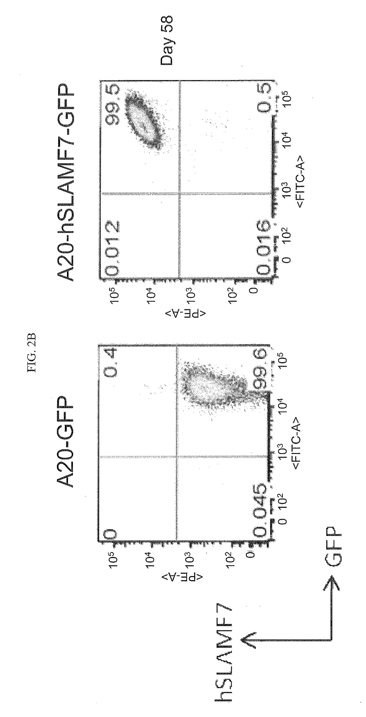
[0247]The A20 mouse B lymphoma cell line was transduced with either retrovirus encoding GFP alone or with retrovirus encoding both GFP and hSLAMF7. A20-GFP and A20-hSLAMF7-GFP lines were sub-cloned, individual clones were picked and expanded in vitro. A20-GFP (clone D3) and A20-hSLAMF7-GFP (clone F11) were maintained in culture and expression of hSLAMF7 and GFP were assessed on day 58 to confirm the stability of hSLAMF7 expression.

[0248]Cells were stained with PE-conjugated anti-human SLAMF7 (clone 162.1, BioLegend) and the frequency of cells staining positive for GFP and hSLAMF7 was determined. As shown in FIGS. 2A-B, A20 cell lines that stably express GFP and hSLAMF7 were obtained.

example 3

Method for Determining Whether Elotuzumab Binds to Human SLAMF7 Expressed in A20 Cells

[0249]To determine whether hSLAMF7 expressed in A20 is recognized by Elotuzumab, A20-GFP and A20-hSLAMF7-GFP cells were stained with Elotuzumab. A20-GFP or A20-hSLAMF7-GFP cells were incubated with 6.25 ug / ml Elotuzumab (BMS), washed twice and incubated with anti-human IgG-PE secondary antibody. The frequency of cells staining positive for GFP and hSLAMF7 was determined using flow cytometry.

[0250]Surface staining, indicating Elotuzumab binding, was detected only in A20-hSLAMF7-GFP cells and not in A20-GFP cells as shown in FIG. 3.


PUM

PropertyMeasurementUnit
timeaaaaaaaaaa
timeaaaaaaaaaa
volumeaaaaaaaaaa

Abstract

The invention described herein relates to therapeutic dosing regimens and combinations thereof for use in enhancing the therapeutic efficacy of anti-CS1 antibodies in combination with an anti-Programmed Death-1 (PD-1) antibody.

Description

[0001]This application claims benefit to provisional application U.S. Ser. No. 62 / 087,489 filed Dec. 4, 2014 under 35 U.S.C. §119(e). The entire teachings of the referenced application are incorporated herein by reference.FIELD OF THE INVENTION[0002]The invention described herein relates to therapeutic dosing regimens and combinations thereof for use in enhancing the therapeutic efficacy of anti-CS1 antibodies in combination with an anti-Programmed Death-1 (PD-1) antibody.BACKGROUND OF THE INVENTION[0003]The National Cancer Institute has estimated that in the United States alone, 1 in 3 people will be struck with cancer during their lifetime. Moreover, approximately 50% to 60% of people contracting cancer will eventually succumb to the disease. The widespread occurrence of this disease underscores the need for improved anticancer regimens for the treatment of malignancy.[0004]Cancer can occur in any tissue or organ of the body. Plasma cell neoplasms, including multiple myeloma, “Sol...

Claims


Application Information

Patent Timeline
Patent Type & Authority Applications(United States)
IPC IPC(8): C07K16/28A61K39/00
CPCC07K16/2818C07K16/2803C07K2317/73C07K2317/21C07K2317/24A61K2039/507A61K2039/545C07K2317/52C07K2317/94C07K2317/76A61K2039/505A61P35/00A61P35/02A61P43/00
Inventor ROBBINS, MICHAEL DARRONGRAZIANO, ROBERT F.BEZMAN, NATALIE
Owner BRISTOL MYERS SQUIBB CO
Why Eureka
Try Eureka