Method and system for improved classification of constituent materials

a constituent material and classification technology, applied in the field of diagnostic imaging, can solve the problems of affecting the quality of the image, the inability of conventional mri to generate response signals with such distinctive signal intensities, and the inability to detect the presence of a single constituent material in the image,

Active Publication Date: 2017-12-28
GENERAL ELECTRIC CO
View PDF0 Cites 10 Cited by
  • Summary
  • Abstract
  • Description
  • Claims
  • Application Information

AI Technical Summary

Problems solved by technology

Typically, PET imaging entails photon-electron interactions that may result in attenuation of emitted photons, which in turn, leads to degraded image quality and inaccurate PET quantitation.
However, conventional MRI fails to generate response signals having such distinctive signal intensities when imaging regions that are devoid of water or fat molecules.
Moreover, MRI of regions proximal to metal objects such as pacemakers or dental fillings may cause excessive heating and / or image artifacts due to large susceptibility variations between metal and surrounding tissue.
Specifically, presence of metals in an imaged region may cause significant resonant frequency changes during MRI, thereby resulting in substantial signal loss, failure of fat suppression, geometric distortion, and bright pile-up artifacts.
However, even such conventional classification methods fail to mitigate inaccuracies in determined diagnostic information resulting from large signal losses caused by presence of metal objects in the imaged region.
Specifically, magnetic susceptibility artifacts that arise due to presence of metal, for example, in the dental fillings or in hip-joint implants distort the diagnostic information derived from MR images.
The distorted MR image information fails to aid in accurate and efficient classification of imaged regions in and / or near air, bones and / or metal.
The inefficient classification, in turn, precludes use of the MR image information for accurate estimation of PET attenuation values and / or any other biochemical investigation.
Magnetic susceptibility artifacts, thus, render conventional MRI unsuitable for a variety of clinical applications.

Method used


Image

  • Method and system for improved classification of constituent materials
  • Method and system for improved classification of constituent materials
  • Method and system for improved classification of constituent materials

Examples


Embodiment Construction

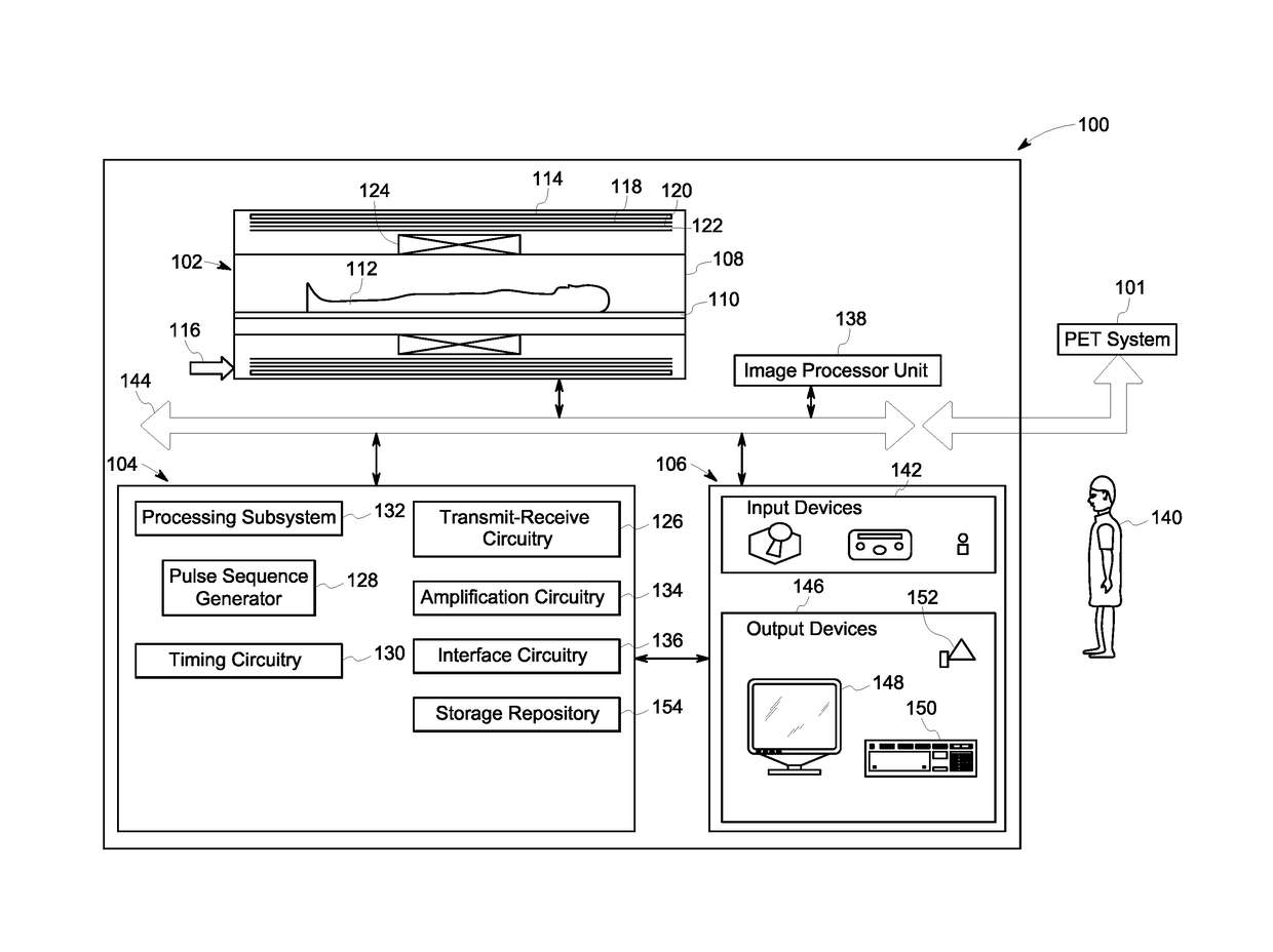
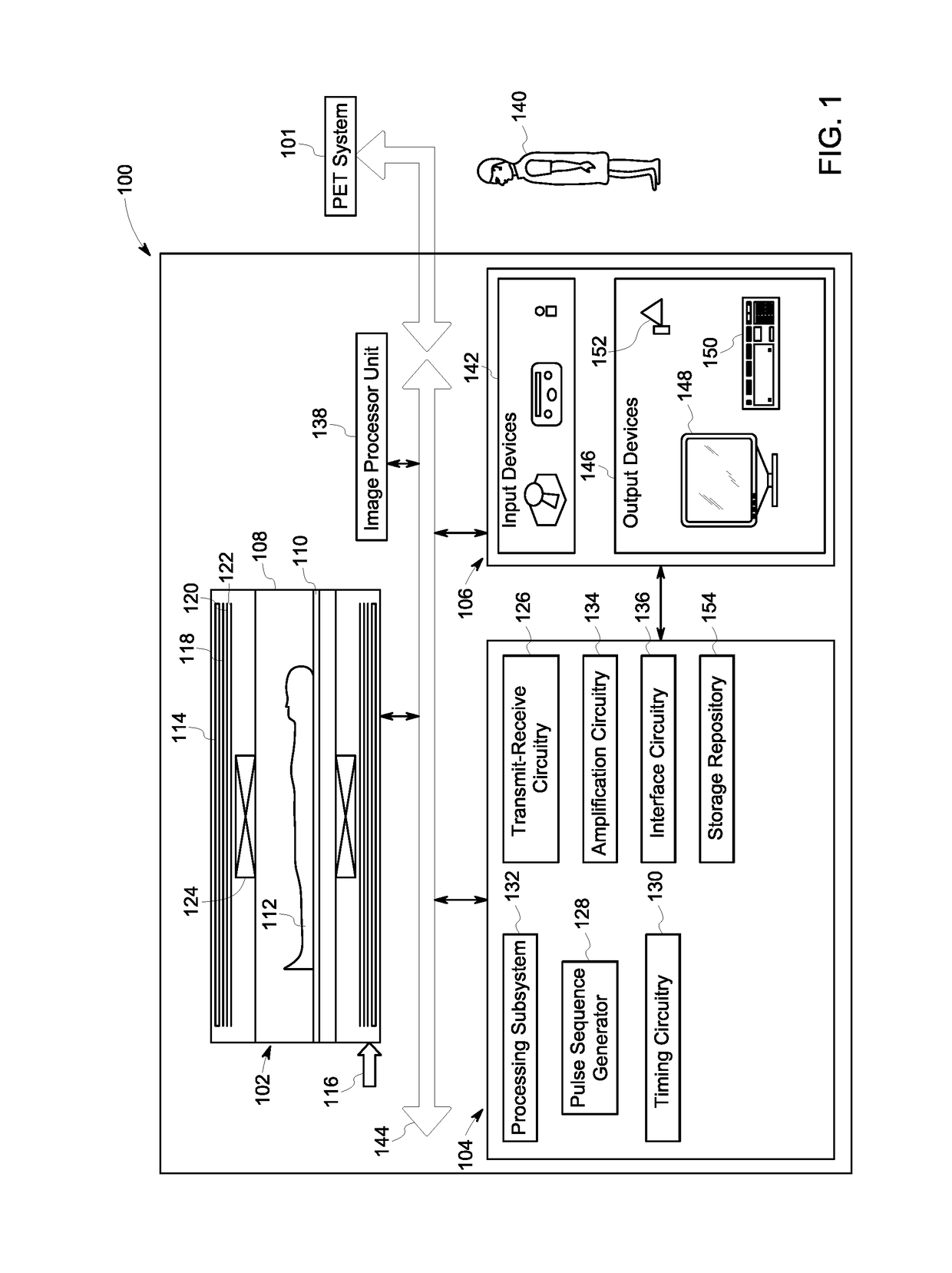
[0017]The following description presents a system and method for accurately classifying constituent materials in a target volume of a subject. Specifically, embodiments described herein allow for accurate identification of constituent materials that are traditionally indistinguishable using conventional magnetic resonance imaging (MRI). In particular, embodiments of the present system and method provide an improved MRI workflow to differentiate a metal-induced signal loss from a signal loss caused by air and / or bone, thereby localizing and / or identifying metal objects located in the target volume with greater accuracy.

[0018]According to certain aspects of the present specification, regions including metal objects may be differentiated from air and / or bone using a combination of magnitude and phase images obtained from acquired MRI data and / or B0 field maps. It may be noted that a B0 field map corresponds to a map of off-resonance frequencies that are generated due to inhomogeneity i...


PUM


Abstract

An imaging system and method are disclosed. An MR image and measured B0 field map of a target volume in a subject are reconstructed, where the MR image includes one or more bright and / or dark regions. One or more distinctive constituent materials corresponding to the bright regions are identified. Each dark region is iteratively labeled as one or more ambiguous constituent materials. Susceptibility values corresponding to each distinctive and iteratively labeled ambiguous constituent material is assigned. A simulated B0 field map is iteratively generated based on the assigned susceptibility values. A similarity metric is determined between the measured and simulated B0 field maps. Constituent materials are identified in the dark regions based on the similarity metric to ascertain corresponding susceptibility values. The MRI data is corrected based on the assigned and ascertained susceptibility values. A diagnostic assessment of the target volume is determined based on the corrected MRI data.

Description

CROSS-REFERENCE TO RELATED APPLICATIONS[0001]This is a national stage application under 35 U.S.C. §371 (c) of PCT Patent Application No. PCT / US2015 / 063392, filed on Dec. 2, 2015, which claims priority to India Patent Application No. 6113 / CHE / 2014, filed on Dec. 4, 2014, the disclosures of which are incorporated herein by reference in their entireties.BACKGROUND[0002]Embodiments of the present specification relate generally to diagnostic imaging, and more particularly to methods and systems for improved classification of constituent materials in a magnetic resonance (MR) image.[0003]Magnetic resonance imaging (MRI) provides high-quality images with excellent soft-tissue contrast for use in diagnosis and / or treatment of a patient. Particularly, MRI may be used in conjunction with one or more other imaging modalities to provide complementary diagnostic information for use in studying biochemical processes in greater detail. For example, MRI may be used to complement molecular informati...

Claims


Application Information

Patent Timeline
Patent Type & Authority Applications(United States)
IPC IPC(8): G01R33/24A61B5/055A61B5/00
CPCG01R33/243A61B5/0035A61B5/055G01R33/481G01R33/4816G01R33/5608G01R33/56536
Inventor SHANBHAG, DATTESH DAYANANDMULLICK, RAKESHTHIRUVENKADAM, SHESHADRIWIESINGER, FLORIANCHATTERJEE, SUDHANYAKOCH, KEVIN MATTHEW
Owner GENERAL ELECTRIC CO
Why Eureka
Try Eureka