Cytologic diagnosis support apparatus, cytologic diagnosis support method, remote diagnosis support system, service providing system, and image processing method

Active Publication Date: 2018-02-22
HITACHI HIGH-TECH CORP
View PDF4 Cites 9 Cited by
  • Summary
  • Abstract
  • Description
  • Claims
  • Application Information

AI Technical Summary

Benefits of technology

[0010]According to the present invention, even in a case where tissues or cells have different shapes in accordance with the type of abnormal cells (for example, cancer) or the degree of progress of abnormal cells (for example, cancer), by

Problems solved by technology

In the pathological diagnosis, the process from specimen preparation to diagnosis requires a lot of manpower, and automation is difficult.
Me

Method used


Image

  • Cytologic diagnosis support apparatus, cytologic diagnosis support method, remote diagnosis support system, service providing system, and image processing method
  • Cytologic diagnosis support apparatus, cytologic diagnosis support method, remote diagnosis support system, service providing system, and image processing method
  • Cytologic diagnosis support apparatus, cytologic diagnosis support method, remote diagnosis support system, service providing system, and image processing method

Examples


first embodiment

(1) First Embodiment

[0031]

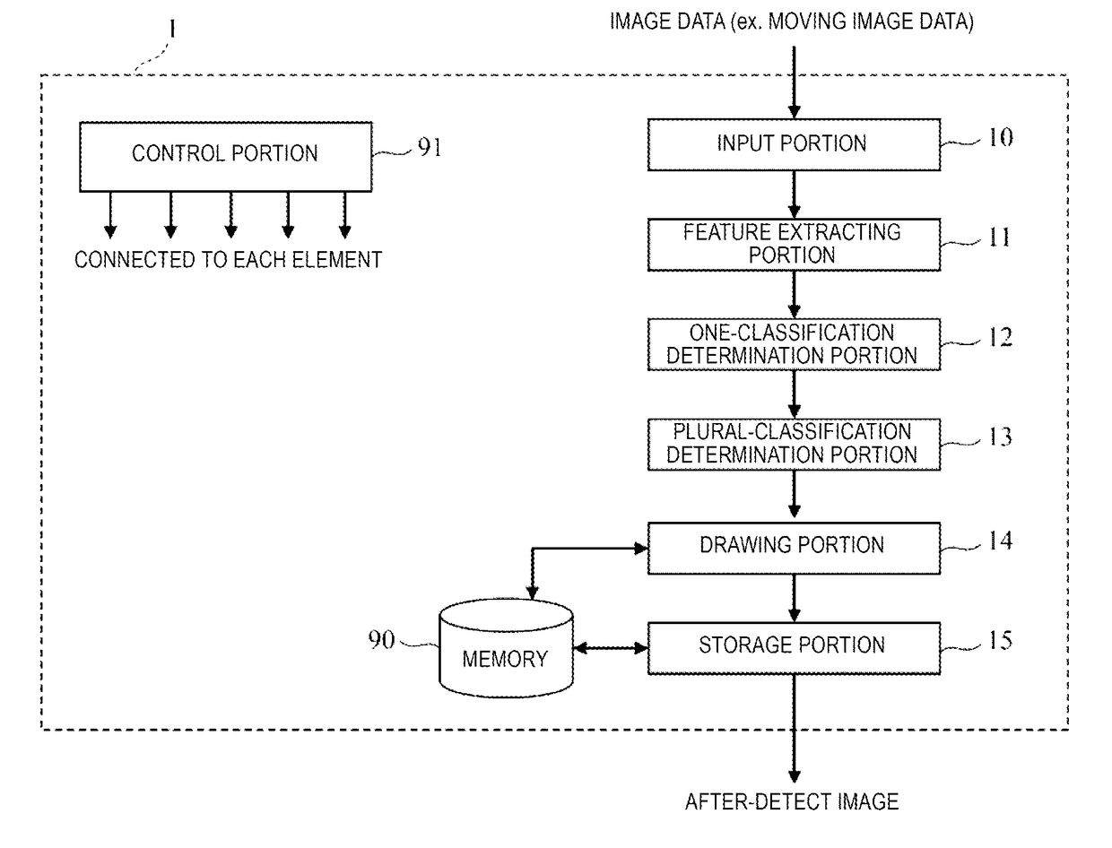
[0032]FIG. 1 is a block diagram illustrating a functional configuration of an image processing apparatus according to the embodiment of the present invention. An image processing apparatus 1 includes an input portion 10, a feature extracting portion 11, a one-classification determination portion 12, a plural-classification determination portion 13, a drawing portion 14, a storage portion 15, a control portion 91, and a memory 90. The image processing apparatus may be mounted in a tissues and cells image obtaining apparatus, such as a virtual slide, and as will be described later (third and fourth embodiments), may be mounted in a server connected to the tissues and cells image obtaining apparatus via a network.

[0033]In the image processing apparatus 1, the input portion 10, the feature extracting portion 11, the one-classification determination portion 12, the plural-classification determination portion 13, the drawing portion 14, and the storage portion 15...

second embodiment

(2) Second Embodiment

[0088]The image processing apparatus 1 according to the second embodiment will be described in FIG. 10. As illustrated in FIG. 10, configurations which are the same as those of FIG. 1 of the first embodiment are included, but the operations of the feature extracting portion 11 and the one-classification determination portion 12 are different from those of FIG. 1. In addition, the learning portion 16 is added. Therefore, here, configurations having different processing and additional configurations will be described by using FIG. 10, and the entire processing flow different from that of FIG. 9 will be described by using FIG. 13.

[0089]

[0090]Hereinafter, configurations and operations of each element different from those of FIG. 1 will be described in detail.

[0091](i) Learning Portion 16

[0092]The learning portion 16 includes the same configuration as that of the feature extracting portion 11 and the one-classification determination portion 12 on the inside thereof, ...

third embodiment

(3) Third Embodiment

[0134]FIG. 14 is a functional block diagram illustrating a configuration of a remote diagnosis support system 1400 according to a third embodiment of the present invention. The remote diagnosis support system 1400 includes a server 1403 and an image obtaining apparatus 1405.

[0135]The image obtaining apparatus 1405 is an apparatus, such as a virtual slide apparatus or a personal computer equipped with a camera, and includes a capturing portion 1401 which captures the image data, and a display portion 1404 for displaying the determination result which has been transmitted from the server or the like 1403. In addition, although not being illustrated, the image obtaining apparatus 1405 includes a communication device which sends the image data to the server or the like 1403 and receives the data that has been sent from the server or the like 1403.

[0136]The server or the like 1403 includes the image processing apparatus 1 which performs the image processing according ...


PUM


Abstract

The present invention realizes determination of tissues and cells from an image by calculating feature values of a deformed degree of the cells even in a case where the tissues or the cells have various shapes. An image diagnosis support apparatus according to the present invention executes processing of inputting an image of cells, processing of extracting the feature values of a plurality of direction components from a target image of the processing, processing of determining whether or not the image corresponds to one-classification by using the plurality of feature values, and processing of determining whether or not the determination processing is finished with respect to all of the classifications set in advance (FIG. 1).

Description

TECHNICAL FIELD[0001]The present invention relates to a cytologic diagnosis support apparatus, a cytologic diagnosis support method, a remote diagnosis support system, a service providing system, and an image processing method, and for example, to an image processing technology for supporting the cytologic diagnosis.BACKGROUND ART[0002]In recent years, in the diagnosis of illness, “pathological diagnosis” using microscopic observation of tissue preparation of a lesioned part occupies a significant position. In the pathological diagnosis, the process from specimen preparation to diagnosis requires a lot of manpower, and automation is difficult. In particular, ability and experiment of a pathologist are important in diagnosis, and the diagnosis depends on personal ability of the pathologist. Meanwhile, the number of cancer patients increases due to population aging, and the number of pathologists is not sufficient at a medical site. From above, needs for image processing technology or...

Claims


Application Information

Patent Timeline
IPC IPC(8): G06T7/00G01N33/483G06K9/46G06K9/62
CPCG06T7/0012G01N33/4833G06K9/4604G06K9/6267G06T2207/30096G06T2207/10088G06T2207/10132G06T2207/10056G06T2207/20081G06T2207/30024G06F18/24
Inventor HATTORI, HIDEHARUKAKISHITA, YASUKIUCHIDA, KENKOASO, SADAMITSUHAMAZATO, FUMIAKI
Owner HITACHI HIGH-TECH CORP
Why Eureka
Try Eureka