Accurate detection and assessment of radiation induced lung injury based on a computational model and computed tomography imaging

Active Publication Date: 2018-03-15
UNIV OF LOUISVILLE RES FOUND INC
View PDF8 Cites 21 Cited by
  • Summary
  • Abstract
  • Description
  • Claims
  • Application Information

AI Technical Summary

Benefits of technology

[0007]A system and computation method is disclosed that overcomes problems associated with conventional a

Problems solved by technology

Although higher radiation doses increase the radiation therapy effectiveness for tumor control, such higher doses may lead to lung injury because, under such conditions, a greater quantity of normal lung tissue may be included in treated areas.
Conventional approaches to the detection and characterization of radiation-induced lung injury are expensive and rely on slow machines that produce images that have insufficient resolution.
Conventional approaches that rely only on the appearance of computed tomography (CT) scans (i.e., Hounsfield Units) do not enable early detection of radiation-induced lung injury, making treatment more difficult.
Although global pulmonary function tests (PFT), such as spirometry, measure air flow obstruction/restriction, no regional lung function information is obtained.
However, SPECT image acquis

Method used


Image

  • Accurate detection and assessment of radiation induced lung injury based on a computational model and computed tomography imaging
  • Accurate detection and assessment of radiation induced lung injury based on a computational model and computed tomography imaging
  • Accurate detection and assessment of radiation induced lung injury based on a computational model and computed tomography imaging

Examples


Embodiment Construction

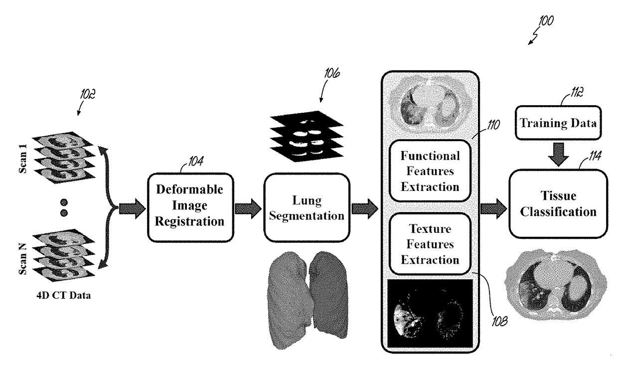
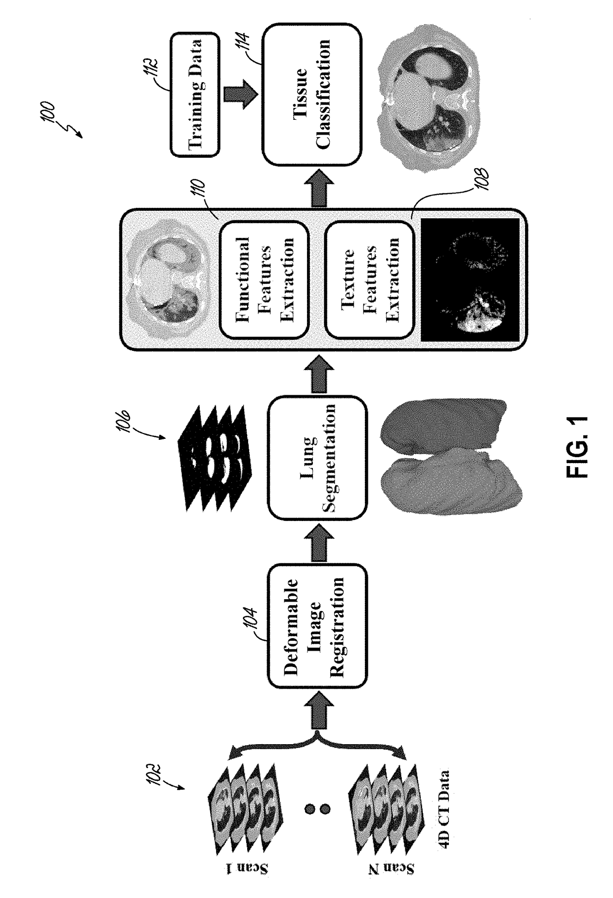
[0051]This document discloses a new and efficient computational framework to accurately align and segment lung regions from four-dimensional computed tomography (4D-CT) images, to extract discriminative features for early detection of radiation-induced lung injury, and to perform the detection.

[0052]FIG. 1 is a schematic illustration of a computational framework 100 that performs efficient detection radiation-induced lung injury, according to an embodiment. The disclosed framework 100, sequentially performs 102 deformable image registration (DIR) of four-dimensional (4D) lung computed tomography (CT) data received as input, performs 106 segmentation of the lung fields with a newly developed image model and methodology, extracts textural 108 and functional 110 features, and performs 114 tissue classification to detect the radiation-induced lung injury using a trainable classifier of lung tissues based on training data 112. The disclosed framework is described in greater detail below....


PUM


Abstract

A system and computation method is disclosed that identifies radiation-induced lung injury after radiation therapy using 4D computed tomography (CT) scans. After deformable image registration, the method segments lung fields, extracts functional and textural features, and classifies lung tissues. The deformable registration locally aligns consecutive phases of the respiratory cycle using gradient descent minimization of the conventional dissimilarity metric. Then an adaptive shape prior, a first-order intensity model, and a second-order lung tissues homogeneity descriptor are integrated to segment the lung fields. In addition to common lung functionality features, such as ventilation and elasticity, specific regional textural features are estimated by modeling the segmented images as samples of a novel 7th-order contrast-offset-invariant Markov-Gibbs random field (MGRF). Finally, a tissue classifier is applied to distinguish between the injured and normal lung tissues.

Description

CROSS REFERENCE TO RELATED APPLICATION[0001]Pursuant to 37 C.F.R. § 1.78(a)(4), this application claims the benefit of and priority to prior filed co-pending Provisional Application Ser. No. 62 / 394,315, filed Sep. 14, 2018, which is expressly incorporated herein by reference in its entirety.FIELD OF THE INVENTION[0002]This disclosure generally relates to the detection and characterization of radiation induced lung injury associated with radiation therapy used to treat lung cancer.BACKGROUND OF THE INVENTION[0003]Radiation-induced lung injury is a significant side effect of radiation therapy for many lung cancer patients. Although higher radiation doses increase the radiation therapy effectiveness for tumor control, such higher doses may lead to lung injury because, under such conditions, a greater quantity of normal lung tissue may be included in treated areas. In recent studies, nearly 40% of patients who underwent radiation therapy developed lung injuries following treatment. Lung...

Claims


Application Information

Patent Timeline
IPC IPC(8): A61B6/00A61B6/03A61N5/10G06T7/33G06T7/149G06T7/143G06T7/174G06T7/38G06T7/246
CPCA61B6/5217A61B6/032A61B6/50A61B6/5235A61N5/1048G06T7/344G06T7/149G06T7/143G06T7/174G06T7/38G06T7/246A61B6/486A61B5/08A61B5/7267G06T2207/10076G06T2207/30061G06T2207/20084G06T2207/20081G06T2207/20076A61B2576/00G06T7/0016G06T2200/04G06T2207/10081G16H50/30
Inventor EL-BAZ, AYMAN S.SOLIMAN, AHMEDKHALIFA, FAHMISHAFFIE, AHMEDDUNLAP, NEALWANG, BRIAN
Owner UNIV OF LOUISVILLE RES FOUND INC
Why Eureka
Try Eureka