Compositions and methods for use of anion channel rhodopsins

a technology anion channel rhodopsin, which is applied in the field of anion channel rhodopsin, can solve the problems of limited photosuppression of neuronal action potentials, and achieve the effects of high-sensitivity and efficient membrane hyperpolarization and neuronal, fast kinetics, and hyperpolarization of the membran

Active Publication Date: 2018-05-03
BOARD OF RGT THE UNIV OF TEXAS SYST
View PDF1 Cites 1 Cited by
  • Summary
  • Abstract
  • Description
  • Claims
  • Application Information

AI Technical Summary

Benefits of technology

[0009]The presently disclosed methods and compositions are based, in part, on the discovery and identification of a novel class of channelrhodopsins, Anion Channel Rhodopsins (ACRs). Light-gated rhodopsin cation channels from chlorophyte algae have transformed neuroscience research through their use as membrane-depolarizing optogenetic tools for targeted photoactivation of neuron firing. Photosuppression of neuronal action potentials has been limited by the lack of equally efficient tools for membrane hyperpolarization. Described herein are Anion Channel Rhodopsins (ACRs), a new family of light-gated anion channels that provide highly sensitive and efficient membrane hyperpolarization and neuronal silencing through light-gated chloride conduction. ACRs strictly conduct anions, completely excluding protons and larger cations, and hyperpolarize the membrane with 100-fold faster kinetics at 3000-fold lower light intensity than the most efficient currently available optogenetic proteins. Sequences encoding 7TM domains of G. theta opsins (295, 291 and 288 aa, corresponding to the JGI protein models 111593, 146828 and 161302, respectively) were optimized for human codon usage and were synthesized. The sequence information encoding the functional constructs 111593 (GtACR1: SEQ ID NO 1) and 146828 (GtACR2: SEQ ID NO: 3) will be represented in GenBank (accession numbers KP171708 and KP171709, respectively). Illumination of neurons expressing GtACR2 fully inhibited their action potential spikes. These ACRs provide new membrane-hyperpolarizing tools for use in establishing a high level of membrane potential for use as optogenetic tools for neuronal silencing of excited cells for among others, neuronal or neurologic disorders, such as but not limited to Parkinson's disease and epilepsy, as well as for cardiac disorders. Thus, ACRs provided herein can serve as new membrane-hyperpolarizing tools for use treatment of epilepsy, as well as for cardiac disorders.

Problems solved by technology

Photosuppression of neuronal action potentials has been limited by the lack of equally efficient tools for membrane hyperpolarization.

Method used


Image

  • Compositions and methods for use of anion channel rhodopsins
  • Compositions and methods for use of anion channel rhodopsins
  • Compositions and methods for use of anion channel rhodopsins

Examples


example 1

and Methods

[0219]Molecular Biology Methods:

[0220]Sequences encoding 7TM domains of G. theta opsins (295, 291 and 288 aa, corresponding to the JGI protein models 111593, 146828 and 161302, respectively) were optimized for human codon usage and were synthesized (Genewiz, South Plainfield, N.J., USA) and cloned into the mammalian expression vector pcDNA3.1 (Life Technologies, Grand Island, N.Y., USA) in frame with an EYFP tag. The sequence information encoding the functional constructs 111593 (GtACR1) and 146828 (GtACR2) were deposited in GenBank (accession numbers KP171708 and KP171709, respectively). The gene encoding archaerhodopsin-3 (Arch) was kindly provided by Dr. Edward S. Boyden (Massachusetts Institute of Technology, Boston, Mass., USA). Mutations were introduced using a QuikChange XL site-directed mutagenesis kit (Agilent Technologies, Santa Clara, Calif., USA) and verified by DNA sequencing.

[0221]HEK293 Recording Methods:

[0222]HEK293 (human embryonic kidney) cells were tran...

example 2

ization of Rhodopsins

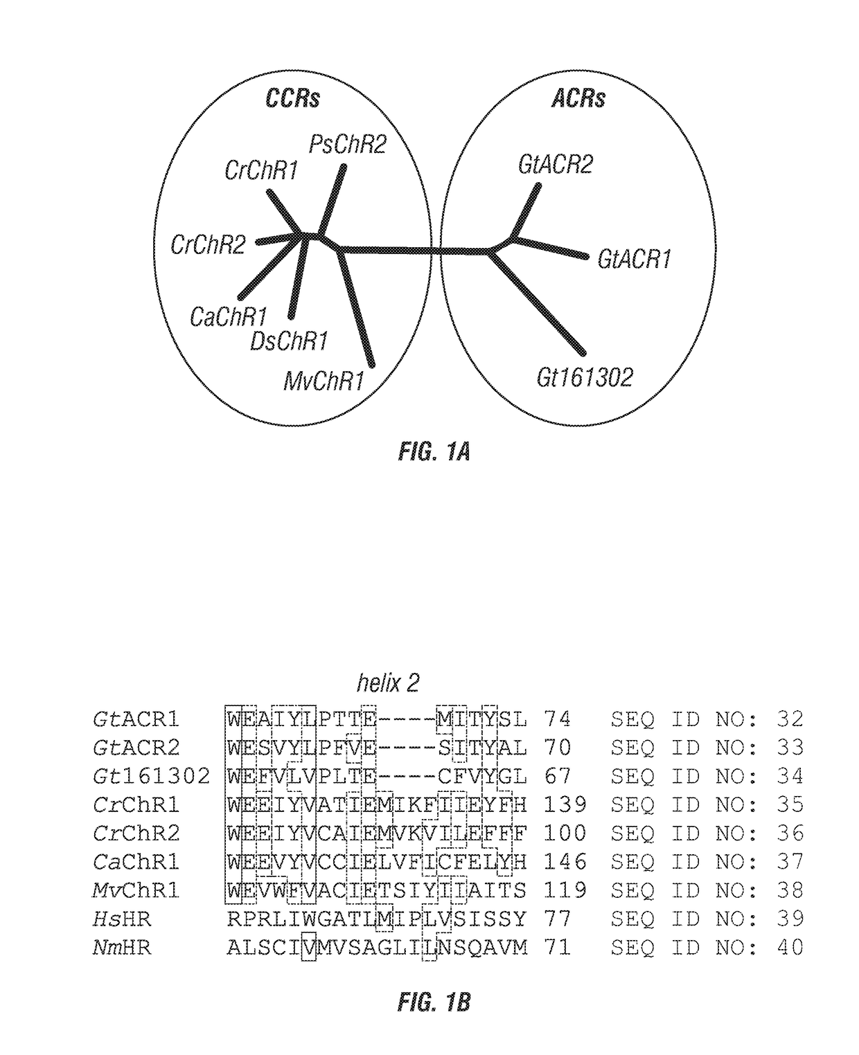
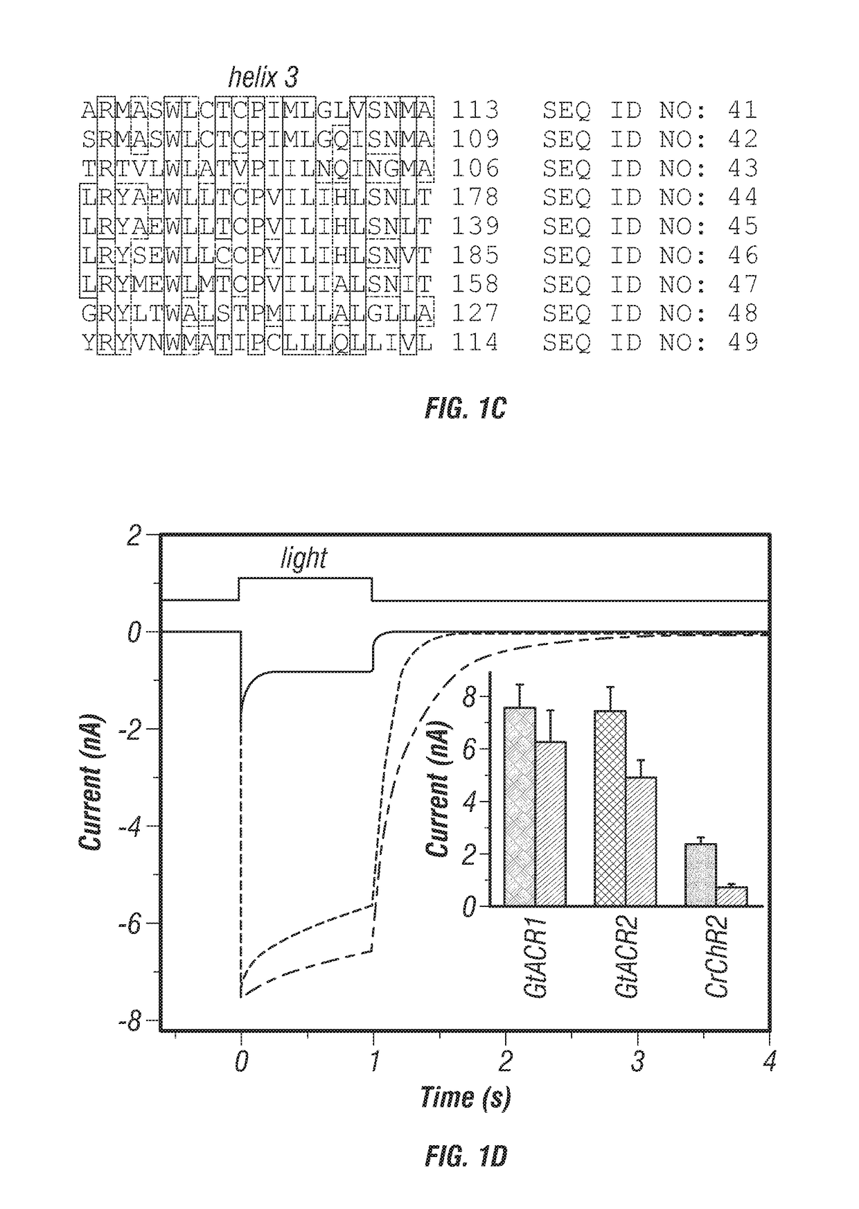
[0226]Of the approximately 50 known ChRs from chlorophytes, all that have been tested are exclusively cation channels. However, several rhodopsin sequences that have been cloned from these organisms did not exhibit channel activity. The nuclear genome sequence of the cryptophyte Guillardia theta has been completely sequenced (B. A. Curtis et al., Nature 492, 59 (2012)). A BLAST search of model proteins returned 53 hits with homology to microbial (type I) rhodopsins. None were highly homologous to ChRs, but the models of one particular cluster (FIG. 1A) do contain some key residues characteristic of chlorophyte ChRs (FIG. 1B).

[0227]The sequences encoding the 7TM domains of G. theta proteins 111593, 146828 and 161302 were well expressed in HEK293 cells. The first two constructs generated photocurrents, whereas the third did not. As shown below, the first two function as light-gated anion channels; therefore we named them GtACR1 and GtACR2 (Guillardia theta Anion C...

example 3

s a Light-Gated Anion Channel (ACR)

[0236]An additional ACR has been identified in the marine cryptophyte Proteomonas sulcata which shows a close similarity to those ACRs described above that were derived from G. theta. This sequence, has been identified as PsChR1 (see GenBank: KF992074, incorporated herein by reference) and has been shown to generate photocurrents when expressed in cultured neurons (Klapoetke N C, Murata Y, Kim S S, Pulver S R, Birdsey-Benson A, Cho Y K, Morimoto T K, Chuong A S, Carpenter E J, Tian Z, Wang J, Xie Y, Yan Z, Zhang Y, Chow B Y, Surek B, Melkonian M, Jayaraman V, Constantine-Paton M, Wong G K, Boyden E S. Nat Methods. 2014 March; 11(3):338-46)), but these photocurrents were not characterized in detail and the measurements did not distinguish between cationic and anionic conductance. In the example below it is demonstrated that the corresponding P. sulcata protein exhibits light-gated anion conductance similar to that of the ACRs from G. theta, although...


PUM

PropertyMeasurementUnit
membrane potentialsaaaaaaaaaa
resistancesaaaaaaaaaa
pHaaaaaaaaaa

Abstract

Methods and compositions used to identify and characterize a new class of rhodopsins derived from algae, which are highly sensitive and efficient anion-conducting channelrhodopsins. The rhodopsin domain of these anion-conducting channelrhodopsins have been cloned and expressed in mammalian systems and thus may be used in, among others, optogenetic applications and as therapeutic agents for electrically active cell mediated disorders.

Description

[0001]This application claims benefit of priority to U.S. Provisional Application Ser. No. 62 / 135,470, filed Mar. 19, 2015, No. 62 / 149,812, filed Apr. 20, 2015, and No. 62 / 261,821, filed Dec. 1, 2015, the entire contents of which are hereby incorporated by reference.[0002]This invention was made with U.S. Government support under Grant Nos. R37GM027750, R21MH098288 and S10RR022531 awarded by the National Institutes of Health. The government has certain rights in the invention.BACKGROUND OF THE INVENTION1. Field of the Invention[0003]This disclosure generally relates to methods and compositions that utilize channelrhodopsins derived from algae, and more particularly to anion-conducting channelrhodopsins which have novel characteristics, for optogenetic applications or for use as therapeutic agents.2. Description of Related Art[0004]Optogenetics (Deisseroth. Nat Methods 8 (1): 26-9, 2011), refers to using optical methods for probing and controlling genetically targeted neurons within ...

Claims


Application Information

Patent Timeline
Patent Type & Authority Applications(United States)
IPC IPC(8): C07K14/405A61K48/00A61P27/02
CPCC07K14/405A61K48/0058A61P27/02A61K38/00A61N5/062C07H21/04C12N15/79C12N2510/00C12N15/74C12N15/82
Inventor SPUDICH, JOHN LEEGOVORUNOVA, ELENA G.SINESHCHEKOV, OLEG A.
Owner BOARD OF RGT THE UNIV OF TEXAS SYST
Why Eureka
Try Eureka