Three-dimensional scanless holographic optogenetics with temporal focusing

a technology of optogenetics and holographic images, applied in the field of three-dimensional scanless holographic optogenetics with temporal focusing, can solve the problems of limited specificity, unsuitable long-term use, and methods developed for optogenetic photostimulation, and achieve high fidelity in space and tim

Active Publication Date: 2019-07-25
RGT UNIV OF CALIFORNIA
View PDF0 Cites 6 Cited by
  • Summary
  • Abstract
  • Description
  • Claims
  • Application Information

AI Technical Summary

Benefits of technology

[0018]The technology is illustrated in the context of two-photon excitation optogenetics applications. Optogenetics requires the ability to activate custom neuron ensembles in vivo and across large volumes of brain tissue by genetically encoding neurons with photosensitive opsins that externally trigger or inhibit action potentials on demand with exposure to precisely focused light. In one preferred embodiment, 3D Scanless Holographic Optogenetics with Temporal focusing (3D-SHOT) apparatus and method allows precise, simultaneous photo-activation of arbitrary sets of neurons anywhere within the addressable volume of the microscope.
[0020]Computer Generated Holography (CGH) is a scanless technique for two-photon optogenetics, which significantly increases temporal precision of photostimulation. The CGH scheme relies on a spatial light modulator (SLM) to distribute a laser beam into multiple targets with custom 3D shapes. Unlike scanning approaches, CGH wide-area holograms matched to the dimensions of each neuron's soma should enable simultaneous, flash-based activation of large numbers of opsin molecules yielding photocurrents with fast kinetics.
[0021]Temporal Focusing (TF) eliminates the trade-off between the target size in the lateral (x,y) plane and axial (z) resolution. TF relies on a diffraction grating placed in the image plane to decompose femtosecond pulses into separate colors, such that the different wavelength components within the original pulse propagate along separate light paths. Each component of the decomposed pulse is broadened in time, which dramatically reduces peak intensity and prevents two-photon absorption until the original pulse constructively interferes at the conjugate image plane of the diffraction grating. This strategy introduces a secondary axial confinement mechanism that restricts the two-photon response to a thin layer around the focal plane. Since the thickness of this layer no longer depends on the dimensions of the targeted area, but rather on the bandwidth of the pulse, this property can be successfully applied for depth-selective two-photon fluorescence imaging and can be been implemented with mechanical scanning and with random-access volume sampling of functional fluorescence.
[0025]For two-photon photostimulation applications, TF activates opsins over a wide area matching the neuron's shape in the focal plane, without compromising depth specificity. TF also mitigates the effects of light scattering even through thick layers of brain tissue.
[0027]The technology naturally expands to photostimulation and ablation surgery for other types of microscopic cells and organisms. Other applications include high speed, scanless 3D photolithography and micro-engraving where the technology can be used to improve spatial resolution, or to accelerate and simplify 3D manufacturing processes.
[0028]As an add-on system in the optical path of a commercial microscope, the technology can enable deep brain photostimulation of single neurons (enhanced spatial resolution) and deep tissue photostimulation of individual cells. Other biological and medical applications include microsurgery with targeted 3D tissue ablation. The technology can also be used as a holographic method for 3D photo lithography and 3D engraving (enhanced engraving resolution and 3D capabilities, with no mechanical parts that is cost effective and faster), or of 3D image display applications.

Problems solved by technology

Electrode array implants may offer good temporal and spatial resolution but are limited to small volumes within the vicinity of the electrode and are not suitable for long-term use because of poor bio-compatibility.
Direct photo-stimulation is possible by wide-field illumination of brain tissue; however, this simple solution activates all the photosensitive neurons simultaneously and offers limited specificity.
In principle, optogenetics offers high spatiotemporal precision, yet the vast majority of optogenetics studies achieve this by genetic specificity rather than spatial control due to the difficulties of accurately focusing light in brain tissue.
Several methods have been developed for optogenetic photostimulation, however none are capable of simultaneous single-neuron spatial resolution across a large volume without compromising temporal precision.
This point-by-point method offers good spatial resolution but poor temporal resolution.
Neurons receive light sequentially and the ability for simultaneous activation of multiple neurons is limited.
This method provides appropriate temporal resolution; however, there is generally a trade-off between operating volume size and spatial resolution.
In both cases, the main limiting factor affecting spatial resolution for photo-stimulation is optical aberration and scattering caused by in-homogeneity of brain tissue, even when using adaptive optics.
However, photosensitive opsins such as ChR2 deactivate rapidly, making it difficult to quickly achieve the photocurrent needed for fast and reliable action potential initiation by activating the opsin point by point in each neuron.
While newly engineered opsins with slow deactivation kinetics may overcome this problem, they necessarily degrade temporal resolution, making it difficult to trigger action potentials with precise timing.

Method used


Image

  • Three-dimensional scanless holographic optogenetics with temporal focusing
  • Three-dimensional scanless holographic optogenetics with temporal focusing
  • Three-dimensional scanless holographic optogenetics with temporal focusing

Examples


example 1

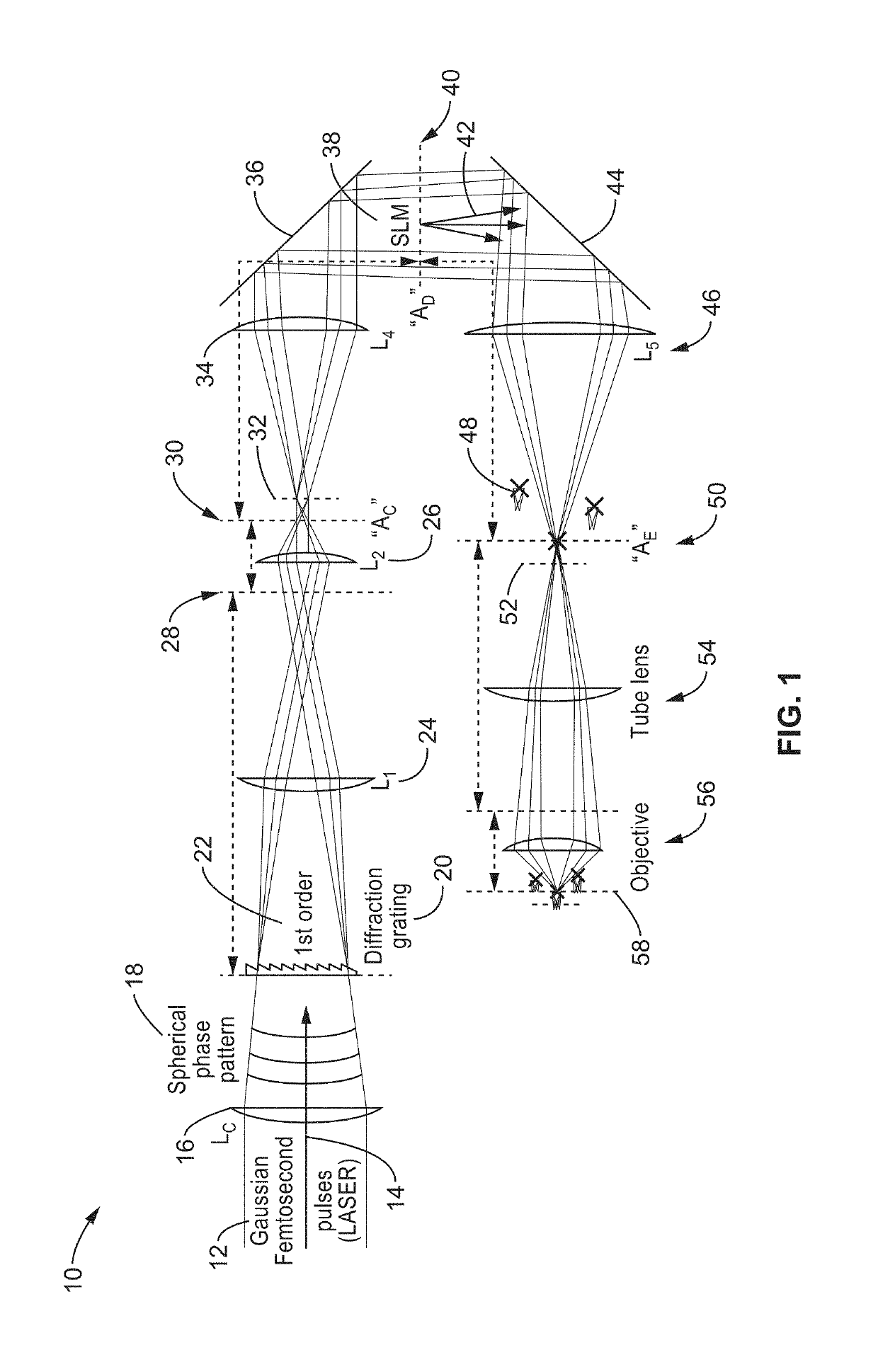
[0081]To evaluate the capabilities of 3D-SHOT and to quantify how two-photon absorption is spatially distributed in 3D, an apparatus was assembled as generally shown in FIG. 1.

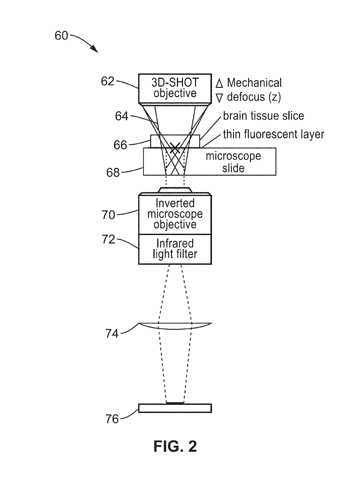
[0082]CGH and 3D-SHOT characterization tests were performed by recording two-photon induced fluorescence on a calibration slide with an inverted microscope. A Basler ACa2500 camera, and a Leitz 6.3× Objective were used to map the entire operational range of the SLM on the camera sensor. Two infrared filters were placed along the light-path to eliminate the remaining laser light, leaving only the fluorescence signal in the visible range to be recorded by the camera sensor.

[0083]For power characterizations, thick auto-fluorescent plastic slides (Chroma) were used to simultaneously collect fluorescence light within the entire volume of excitation from a single focused image. For more precise 3D characterization of two photon absorption, a custom-made thin film of fluorescent paint (Tamiya Color TS-36 fluorescent ...

example 2

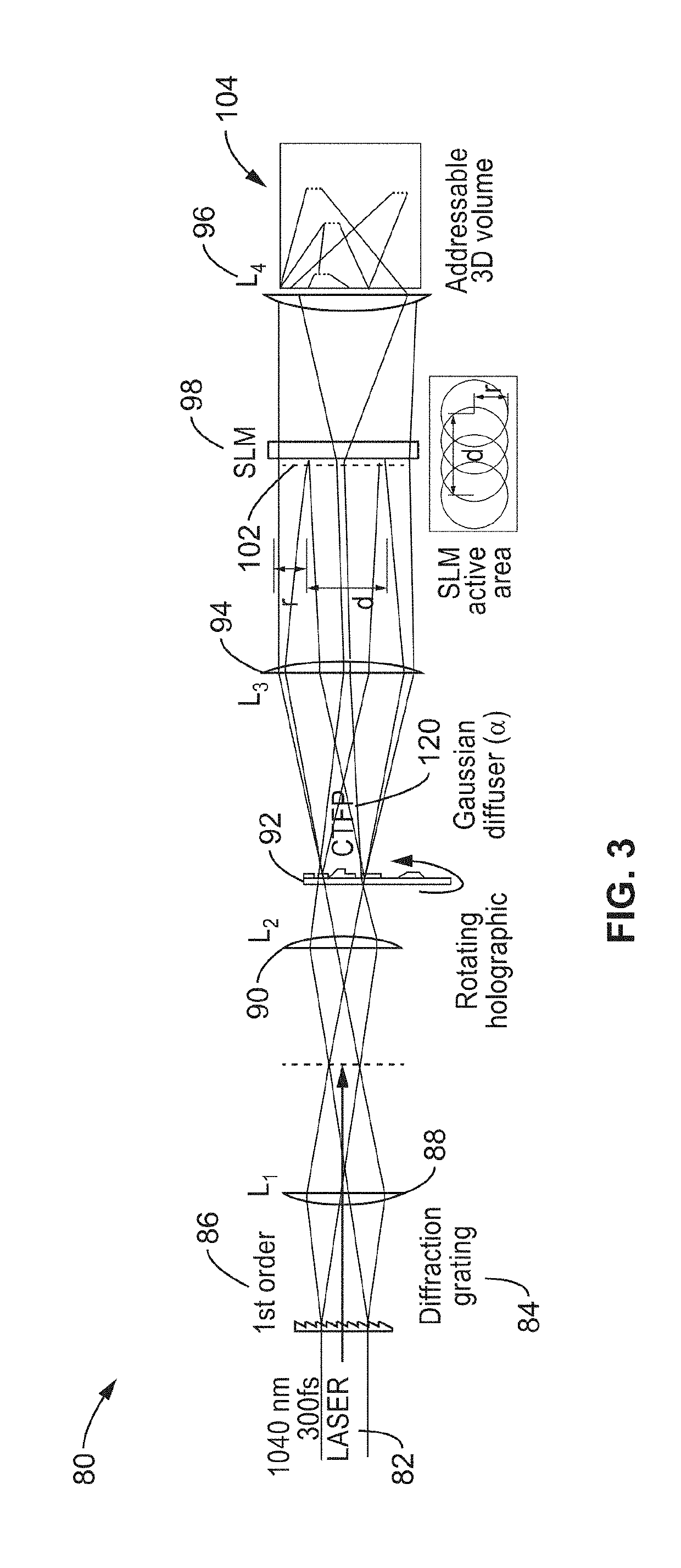
[0093]The 3D-SHOT apparatus and methods were also evaluated for use in neurobiological applications. The apparatus was configured with a pair of mirrors on a sliding stage to rapidly swap between conventional holography (CGH) and 3D-SHOT. The two optical paths were aligned so that the centers of the CTFP (3D-SHOT path) and the disc illumination (CGH path) were co-aligned along all 3 dimensions. This allowed both methods to be tested on individual cells without any digital realignment of the holograms.

[0094]The spatial resolution of the 3D-SHOT method was evaluated with quantitative measurements of the photocurrent amplitudes elicited by optogenetic stimulation in living cells. Microbial opsin proteins were expressed in either Chinese Hamster Ovary (CHO) cells in culture by transfection, or through in utero electroporation of mice embryos or viral transduction of neonatal mice to obtain expression in neurons in acutely prepared cortical brain slices or in vivo.

[0095]To measure the sp...

example 3

[0105]To further demonstrate the functionality of the apparatus and optical methods, new opsins were engineered to be optimized for multiphoton optogenetics using scanless approaches. Existing excitatory opsins have been shown to be too weak or too slow to drive precise neural activity patterns with scanless 2P optogenetics in vivo. To allow control of neural activity with sub-millisecond precision, an opsin and a 2P stimulation approach capable of generating large currents with rapid kinetics was sought. Several conventional opsins were evaluated. However, photocurrents were insufficient to reliably spike pyramidal neurons. Since none of the conventional opsins could reliably spike neurons in response to brief holographic stimulation, a stronger opsin was engineered with the goal of holographically stimulating large ensembles of neurons.

[0106]Mutations of the ST-Chronos opsin were performed with the aim of developing a variant that would preserve its fast kinetics but would generat...


PUM


Abstract

Apparatus and methods for 3D-Scanless Holographic Optogenetics with Temporal focusing (3D-SHOT), which allows precise, simultaneous photo-activation of arbitrary sets of neurons anywhere within the addressable volume of the microscope. Soma-targeted (ST) optogenetic tools, ST-ChroME and IRES-ST-eGtACR 1, optimized for multiphoton activation and suppression are also provided. The methods use point-cloud holography to place multiple copies of a temporally focused disc matching the dimensions of a designated neuron's cell body. Experiments in cultured cells, brain slices, and in living mice demonstrate single-neuron spatial resolution even when optically targeting randomly distributed groups of neurons in 3D.

Description

CROSS-REFERENCE TO RELATED APPLICATIONS[0001]This application claims priority to, and the benefit of, U.S. provisional patent application Ser. No. 62 / 620,601 filed on Jan. 23, 2018, incorporated herein by reference in its entirety.STATEMENT REGARDING FEDERALLY SPONSORED RESEARCH OR DEVELOPMENT[0002]This invention was made with Government support under NS087725 awarded by The National Institutes of Health. The Government has certain rights in the invention.INCORPORATION-BY-REFERENCE OF SEQUENCE LISTING[0003]This application includes a sequence listing in a text file entitled “ST-ChroME-sequence-ST25.txt” created on Jan. 23, 2019 and having a 22 kb file size. The sequence listings are submitted through EFS-Web and are incorporated herein by reference in their entirety.NOTICE OF MATERIAL SUBJECT TO COPYRIGHT PROTECTION[0004]A portion of the material in this patent document is subject to copyright protection under the copyright laws of the United States and of other countries. The owner...

Claims


Application Information

Patent Timeline
Patent Type & Authority Applications(United States)
IPC IPC(8): G03H1/04G03H1/00G06N3/067G03H1/08
CPCG03H1/0443G03H1/0005G06N3/0675G03H1/0866G03H2001/005G03H2001/0883G03H2001/0077G03H2001/2213G03H2001/0216G03H2223/13G03H2223/23G03H2222/16G03H2222/33G03H2210/30G03H2222/14G03H1/2294G03H1/2249G03H1/2286G06N3/061G02B21/06
Inventor WALLER, LAURAADESNIK, HILLELPEGARD, NICOLAS C.
Owner RGT UNIV OF CALIFORNIA
Why Eureka
Try Eureka