Methods and systems for assessing retinal images, and obtaining information from retinal images

a retinal image and information technology, applied in image enhancement, medical/anatomical pattern recognition, instruments, etc., can solve the problems of damage and degeneration of the retinal layer, vision loss is permanent and irrecoverable, and early detection is often further complicated, so as to improve the reliability of existing retinal image processing systems, and capture a good retinal interest region or overall image

Active Publication Date: 2018-06-28
AGENCY FOR SCI TECH & RES
View PDF3 Cites 8 Cited by
  • Summary
  • Abstract
  • Description
  • Claims
  • Application Information

AI Technical Summary

Benefits of technology

[0026]An embodiment of the invention can be easily used in current technologies without the need for extensive modifications. For example, an embodiment can be used in two scenarios.
[0027]1) It can be used as a pre-filter for current automated disease detection systems by assessing the quality of retinal interest regions. Further, by successively applying the embodiment onto subsequent regions, the image can be fully probed to find a useable region instead of rejecting the image based on global quality. The embodiment may ensure that input regions are of usable quality, and provide feedback on the reliability of results bas

Problems solved by technology

However, in many of the other diseases such as glaucoma and AMD, vision loss tends to be permanent and irrecoverable.
Early detection is often further complicated by the asymptomatic nature of many eye diseases, in which visual symptoms are usually only observed by the patient at an advanced stage of progression.
Damage and degeneration of the retinal layer are key causes of permanent visual loss.
However, direct observation of the optic disk using modalities such as retinal imaging can be instructive in glaucoma detection.
In AMD, degeneration of the macular region of the retina leads to a direct loss of vision in the center of the visual field.
However, i

Method used


Image

  • Methods and systems for assessing retinal images, and obtaining information from retinal images
  • Methods and systems for assessing retinal images, and obtaining information from retinal images
  • Methods and systems for assessing retinal images, and obtaining information from retinal images

Examples


Embodiment Construction

[0052]Referring firstly to FIG. 6, a flow diagram is shown of a method 100 which is an embodiment of the method. The embodiment is referred to as ARIES (an Automated Retinal Interest Estimator System). ARIES automatically assesses the quality of input images as a pre-processing step before passing the processed images for subsequent analysis. In this way, ARIES will control the quality of input for subsequent analysis.

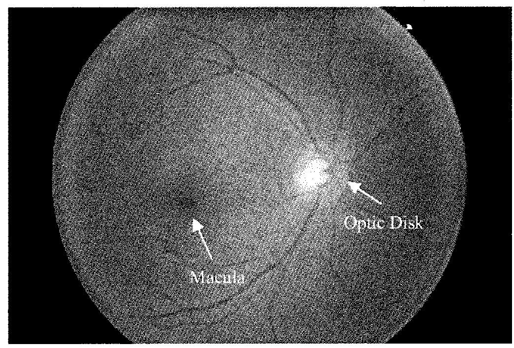
[0053]A key feature of ARIES is that it analyzes at least one of the focal regions of interest. A specific example here is the optic disk. Since imaging artefacts can be local, ARIES will help to ensure that the usage of the input image is maximized by assessing the quality of the detected focal region of interest, rather than rejecting the entire image based on global characteristics. If the initial region of interest is not suitable, another ROI will be extracted and re-assessed. This is repeated until a suitable ROI is found, or when all possible ROI are exhausted. ...


PUM


Abstract

A method of assessing the quality of an retinal image (such as a fundus image) includes selecting at least one region of interest within a retinal image corresponding to a particular structure of the eye (e.g. the optic disc or the macula), and a quality score is calculated in respect of the, or each, region-of-interest. Each region of interest is typically one associated with pathology, as the optic disc and the macula are. Optionally, a quality score may be calculated also in respect of the eye as a whole (i.e. over the entire image, if the entire image corresponds to the retina).

Description

FIELD OF THE INVENTION[0001]The present invention relates to computer-implemented methods and apparatus for assessing images which are believed to be images of the retina of an eye, and, in the case that the images are determined to be of sufficient quality, for obtaining information from them, for suggesting a treatment.BACKGROUND OF THE INVENTION[0002]The major causes of blindness include cataracts, glaucoma, age-related macular degeneration, diabetic retinopathy, as well as pathological myopia. Of these, vision lost in cataracts can usually be restored by replacing the opacified crystalline lens with an artificial intra-operative lens. However, in many of the other diseases such as glaucoma and AMD, vision loss tends to be permanent and irrecoverable. Hence, there is considerable public and clinical interest to detect such diseases in as early a stage as possible, in order to save sight and reduce the costs of treatment. Early detection is often further complicated by the asympto...

Claims


Application Information

Patent Timeline
IPC IPC(8): G06K9/46G06K9/00G06K9/03G06T7/00A61B3/00A61B3/12A61B3/14G06V10/764
CPCG06K9/4676G06K9/0061G06K9/036G06T7/0014A61B3/0025A61B3/12A61B3/14G06K2209/05G06T2207/30041G06T2207/30168A61B2576/02G06T7/0012G06T2207/20081G06V40/193G06V40/14G06V10/993G06V10/464G06V2201/03G06V10/764G06F18/2411
Inventor YIN, FENGSHOUWONG, WING KEE DAMONLIU, JIANGLEE, BENG HAIZHANG, ZHUOGOPALAKRISHNAN, KAVITHAQUAN, YINGYOW, AL PING
Owner AGENCY FOR SCI TECH & RES
Why Eureka
Try Eureka