Methods of reducing corneal endothelial cell loss

a corneal endothelial cell and cell technology, applied in the field of reducing can solve the problems of partial neurotrophic keratopathy, damage to corneal nerves, etc., and achieve the effects of reducing nerve loss-related corneal endothelial cell loss, and reducing the number of corneal nerves

Inactive Publication Date: 2018-07-19
MASSACHUSETTS EYE & EAR INFARY +1
View PDF0 Cites 0 Cited by
  • Summary
  • Abstract
  • Description
  • Claims
  • Application Information

AI Technical Summary

Benefits of technology

[0004]Provided herein are methods of reducing nerve loss-related corneal endothelial cell loss in a subject that include selecting a subject identified as having an eye with reduced numbers of corneal nerves as compared to a reference eye, e.g., an eye of a healthy control, and administering vasoactive intestinal peptide (VIP) to the selected subject. In some embodiments of any of the methods described herein, the VIP is topically administered to the eye of the subject. In some embodiments of any of the methods described herein, the VIP is administered to the eye of the subject by systemic administration, subconjunctival injection, or intraperitoneal injection.
[0008]Also provided herein is a VIP or a nucleic acid encoding a VIP for use in reducing nerve loss-related corneal endothelial cell loss in a subject (e.g., a subject identified as having an eye with reduced numbers of corneal nerves as compared to a reference eye, e.g., an eye of a healthy control).
[0009]Also provided herein are methods of using a VIP or a nucleic acid encoding a VIP in the manufacture of a medicament for reducing nerve loss-related corneal endothelial cell loss in a subject (e.g., a subject idenfitied as having an eye with reduced numbers of corneal nerves as compared to a reference eye, e.g., an eye of a healthy control).
[0011]By the term “treating” or “efficacy of treatment” is meant a reduction in the number of symptoms of a disease or disorder in a subject (e.g., reduce the number of symptoms of Fuchs' endothelial corneal dystrophy, pseudophakic bullous keratopathy, keratoconus, pseudoexfoliation syndrome, atopic keratoconjunctivitis, herpetic keratitis, endothelial cell loss after full-thickness or partial-thickness corneal transplantation, herpes zoster ophthalmicus, uveitis, or graft rejection), a decrease (e.g., a significant, detectable, or observable decrease) the severity, frequency, and / or duration of one or more (e.g., at least two, three, or four) symptoms of a disease or disorder in a subject (e.g., reduce the severity, frequency, and / or duration of one or more symptoms of Fuchs' endothelial corneal dystrophy, pseudophakic bullous keratopathy, keratoconus, pseudoexfoliation syndrome, atopic keratoconjunctivitis, herpetic keratitis, endothelial cell loss after full-thickness or partial-thickness corneal transplantation, herpes zoster ophthalmicus, uveitis, or graft rejection in a subject), and / or a decrease corneal endothelial cell loss (e.g., nerve loss-related corneal endothelial cell loss) in a subject.

Problems solved by technology

Corneal nerves can be damaged due to many pathological conditions, such as, e.g., ocular infection, surgery, diabetes, stroke, dry eye syndrome, and intracranial lesion involving the trigeminal nerve, which all result in complete or partial neurotrophic keratopathy.

Method used


Image

  • Methods of reducing corneal endothelial cell loss
  • Methods of reducing corneal endothelial cell loss
  • Methods of reducing corneal endothelial cell loss

Examples


example

Example 1

Nerve Loss-Related Corneal Endothelial Cell Loss and Ability of VIP to Reduce Nerve Loss-Related Corneal Endothelial Cell Loss

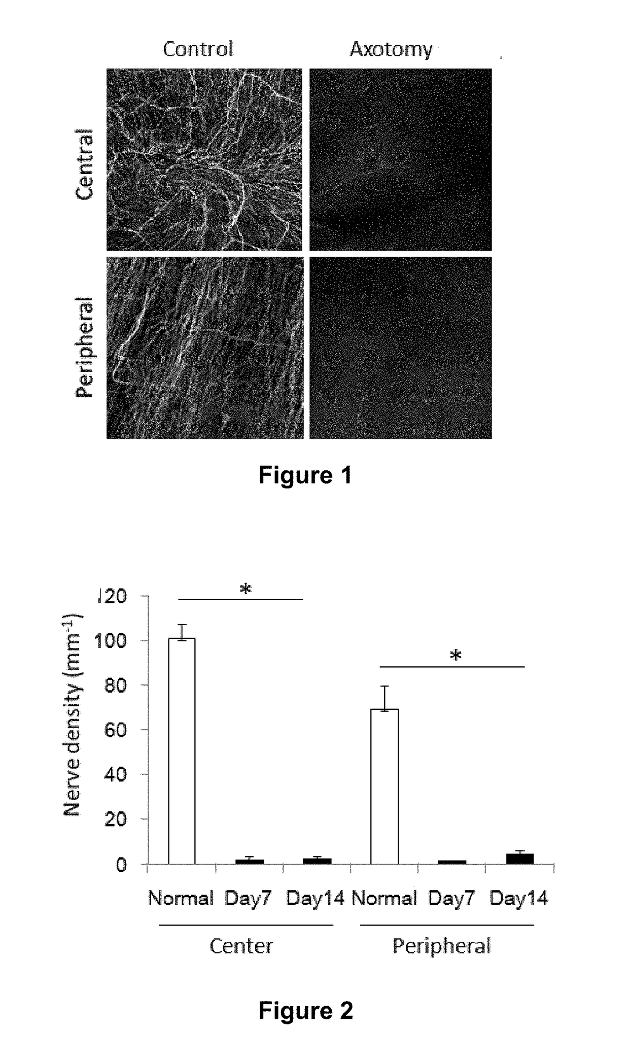
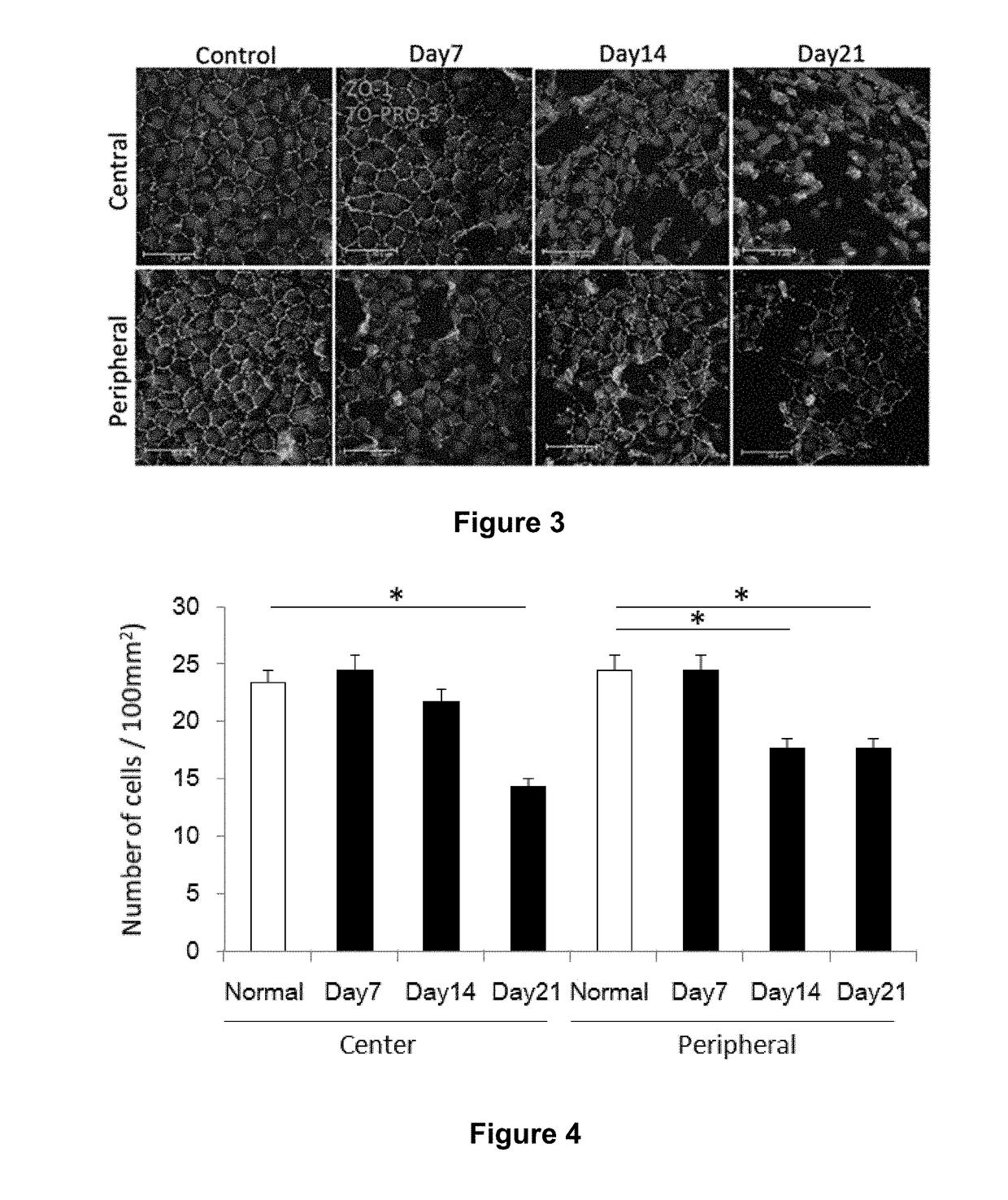
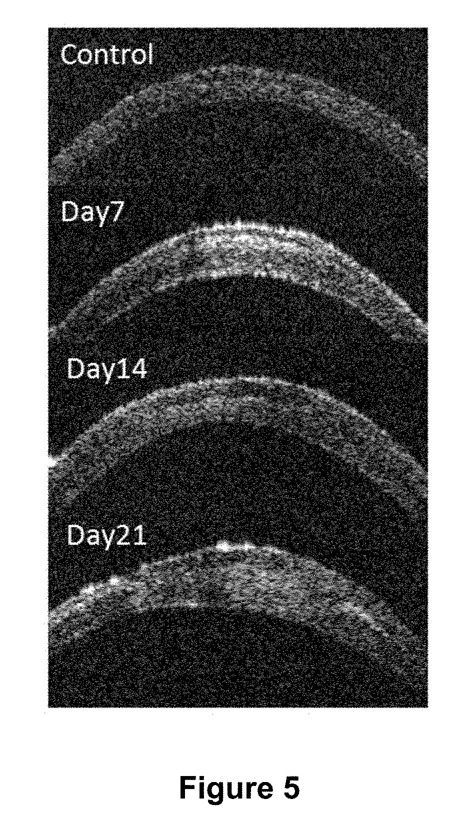
[0101]A set of experiments was performed to evaluate corneal endothelial cell alterations after trigeminal axotomy and the effect of VIP on corneal endothelial cells after trigeminal axotomy.

Materials and Methods

Animals and Surgical Procedure

[0102]Six- to eight-week old male BALB / c mice (Charles River, Wilmington, Mass.) were used in these experiments. Trigeminal axotomy was performed by first anesthetizing the animals with a ketamine (100 mg / mL) / xylazine (20 mg / mL) / acepromazine (15 mg / mL) mixture. After anesthetization, small incision lateral canthotomy was performed, two tractional sutures were placed on the lid skin, and the conjunctival fornix were incised circumferentially around 90 degrees. The eye globe was rotated nasally by gently pushing the nasal fornix with blunt forceps, exposing the trigeminal nerve and minimizing intraoperative bleedin...


PUM

PropertyMeasurementUnit
diameteraaaaaaaaaa
diameteraaaaaaaaaa
diameteraaaaaaaaaa

Abstract

Provided herein are methods of reducing corneal endothelial cell loss (e.g., nerve loss-related corneal endothelial cell loss) that include selecting a subject identified as having an eye with reduced numbers of corneal nerves as compared to a reference eye, e.g., an eye of a healthy control, and administering vasoactive intestinal peptide (VIP) or a nucleic acid encoding VIP to the selected subject.

Description

CROSS-REFERENCE TO RELATED APPLICATIONS[0001]This application is a continuation of U.S. patent application Ser. No. 15 / 037,292, filed on May 17, 2016, which is the U.S. National Stage under 35 USC § 371 of PCT / US2014 / 066551, filed on Nov. 20, 2014, which claims priority to U.S. Provisional Patent Application No. 61 / 906,723, filed on Nov. 20, 2013, the contents of which are incorporated herein by reference in their entirety.BACKGROUND OF THE INVENTION[0002]The cornea is the most densely innervated tissue in the mammalian body. Intact innervation of the cornea is necessary for the maintenance of corneal structure and function (Araki et al., Curr. Eye. Res. 13:203-211, 1994; Nishida et al., Curr. Opin. Ophthalmol. 20:276-281, 2009). Corneal nerves can be damaged due to many pathological conditions, such as, e.g., ocular infection, surgery, diabetes, stroke, dry eye syndrome, and intracranial lesion involving the trigeminal nerve, which all result in complete or partial neurotrophic ker...

Claims


Application Information

Patent Timeline
Patent Type & Authority Applications(United States)
IPC IPC(8): A61K38/22A61K9/00A61B3/10
CPCA61K9/0048A61B3/1025A61K38/2278A61P27/02
Inventor YAMAGUCHI, TAKEFUMIJURKUNAS, ULAHAMRAH, PEDRAM
Owner MASSACHUSETTS EYE & EAR INFARY
Why Eureka
Try Eureka