

Protein-protein interaction detection systems and methods of use thereof
a detection system and protein technology, applied in the field of proteinprotein interaction detection systems, can solve the problems of low sensitivity, lack of temporal control, and requirement for long stimulation periods
- Summary
- Abstract
- Description
- Claims
- Application Information
AI Technical Summary
Benefits of technology
Problems solved by technology
Method used
Image

Examples
example 1
tion Systems
[0514]FIGS. 17-20 provide sequence information regarding exemplary PPI detection systems.
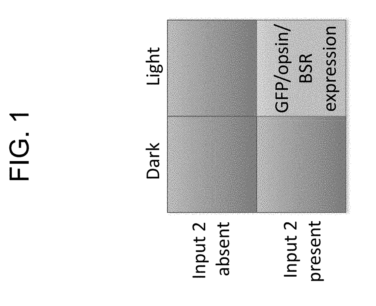
[0515]FIG. 1 is a schematic depiction of the requirement for two input signals for functioning of a system of the present disclosure.
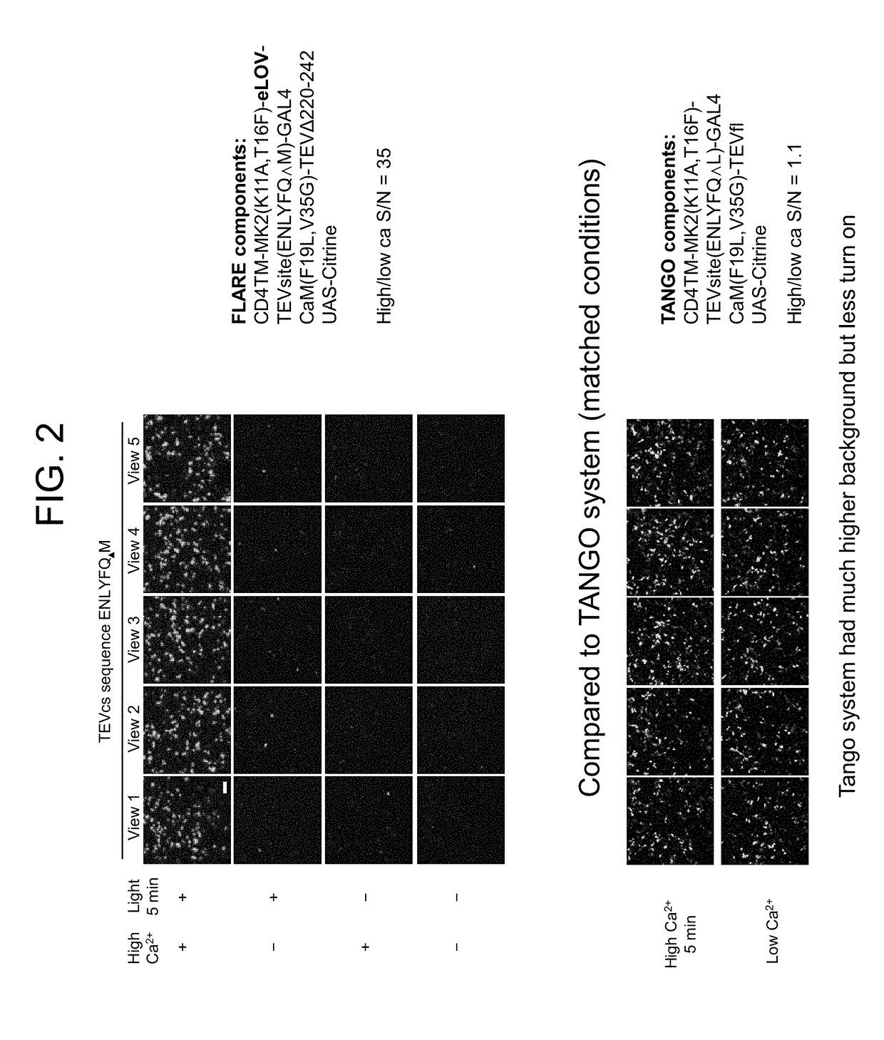
[0516]FIG. 2 presents a comparison of a calcium-induced protein-protein interaction (PPI) detection system of the present disclosure to the TANGO system.
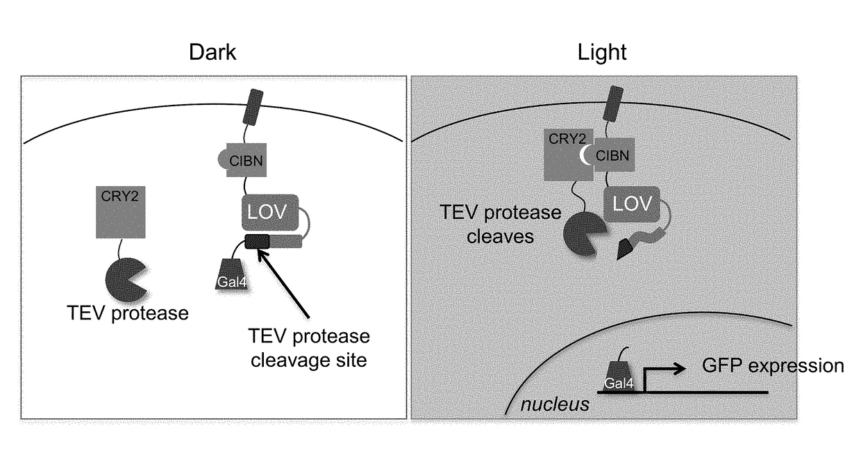
[0517]FIG. 3 is a schematic depiction of an example of a blue light induced CRY2-CIBN PPI detection system.
[0518]FIG. 4 depicts PPI detection using a PPI detection system as schematically depicted in FIG. 3.
[0519]FIG. 5 is a schematic depiction of an isoproterenol induced beta2-AR and beta2-arrestin PPI detection system of the present disclosure.
[0520]FIG. 6 is a workflow diagram for use of a PPI detection system as schematically depicted in FIG. 5.
[0521]FIG. 7 and FIG. 8 depict PPI detection using a PPI detection system as schematically depicted in FIG. 5.
[0522]FIG. 9 is a schematic depiction of a rapa...
example 2
[0524]FIG. 21A-21F: Design of FLARE-PPI and Application to Light- and Agonist-Dependent Detection of β2-Adrenergic Receptor (β2AR)-Arrestin2 Interaction.
[0525](A) Scheme. A and B are proteins that interact under certain conditions. Protein A is membrane-associated and is fused to a light-sensitive eLOV domain, a protease cleavage site (TEVcs), and a transcription factor (TF). These comprise the “FLARE TF component.” Protein B is fused to a truncated variant of TEV protease (TEVp) (“FLARE protease component”). When A and B interact (right), TEVp is recruited to the vicinity of TEVcs. When blue light is applied to the cells, eLOV reversibly unblocks TEVcs. Hence, the coincidence of light and A-B interaction permits cleavage of TEVcs by TEVp, resulting in the release of the TF, which translocates to the nucleus and drives transcription of a reporter gene of interest. (B) FLARE-PPI constructs for studying the β2AR-arrestin interaction. V5 and myc are epitope tags. UAS is a promoter reco...
example 3
on of FLARE-PPI to a Variety of PPIs
[0535]FIG. 27A-27D: FLARE-PPI can be Applied to a Variety of PPIs.
[0536](A) PPI pairs studied with FLARE. DRD1 and NMBR are GPCRs that interact with arrestin2. EGFR is a receptor tyrosine kinase that recruits Grb2 upon stimulation with EGF ligand. FKBP and FRB are soluble proteins that heterodimerize upon addition of the drug rapamycin; to keep FRB FLARE out of the nucleus in the basal state, the FRB-FLARE was fused to either a plasma membrane anchor (TM from CD4) or a mitochondrial membrane anchor (TM from AKAP1). CIBN-CRY2 PHR is a light-inducible PPI. Kennedy et al. (2011) Nat. Methods 7:973-975. (B) FLARE data corresponding to PPIs depicted in (A). FLARE constructs were the same as those shown in FIG. 21B, except β2AR and arrestin2 were replaced by the A and B proteins indicated, respectively. HEK 293T cells transiently expressing FLARE constructs were stimulated with light and the ligand indicated in (A) for 5 minutes, then fixed and imaged 9...
PUM
| Property | Measurement | Unit |
|---|---|---|
| ionic strength | aaaaa | aaaaa |
| ionic strength | aaaaa | aaaaa |
| ionic strength | aaaaa | aaaaa |
Abstract
Description
Claims
Application Information