
Ultrasound compatible x-ray Anti-scatter grid
a detector module and anti-scatter technology, applied in the field of anti-scatter grids, detector modules and imaging apparatuses, can solve the problems of cumbersome multi-modal imaging types in applications and the image quality in such settings remains behind expectations, and achieves the effect of convenient operation of imaging systems
- Summary
- Abstract
- Description
- Claims
- Application Information
AI Technical Summary
Benefits of technology
Problems solved by technology
Method used
Image

Examples
Embodiment Construction
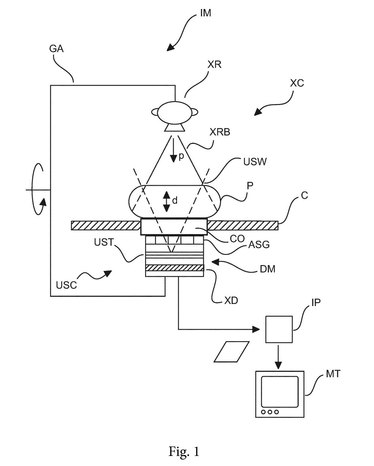
[0045]With reference to FIG. 1, there are shown components of an imaging system IM.
[0046]The imaging system IM is multimodal in the sense that it combines two imaging components that work on different principles: there is an X-ray imaging component XC and there is an ultrasound imaging component USC. Possible, non-exhaustive areas of application include mammography or cardio-vascular imaging.
[0047]Each imaging component can be made to work separately and independent from each other. One component may be used to acquire a first image set and, subsequently thereto, the other component may be used to acquire a second image set. Preferably both components, ultrasound and X-ray, can work concurrently to acquire image sets at the same time of an object to be imaged P. The object is a human or animal (in particular mammal) patient. What is mainly envisaged herein is a human patient or an anatomical part thereof. In the following we will therefore refer to the object as patient P. In one em...
PUM
 Abstract
Description
Claims
Application Information