Retinal stem cells
- Summary
- Abstract
- Description
- Claims
- Application Information
AI Technical Summary
Benefits of technology
Problems solved by technology
Method used
Image

Examples
example 1
[0079]Isolation of retinal stem cells from the ora terminalis of a human eye was performed in the present experiment.
[0080]The ora terminalis was dissociated and cultured using an assay for forming a clonal sphere, in which the stem cells form clonally derived spheres.
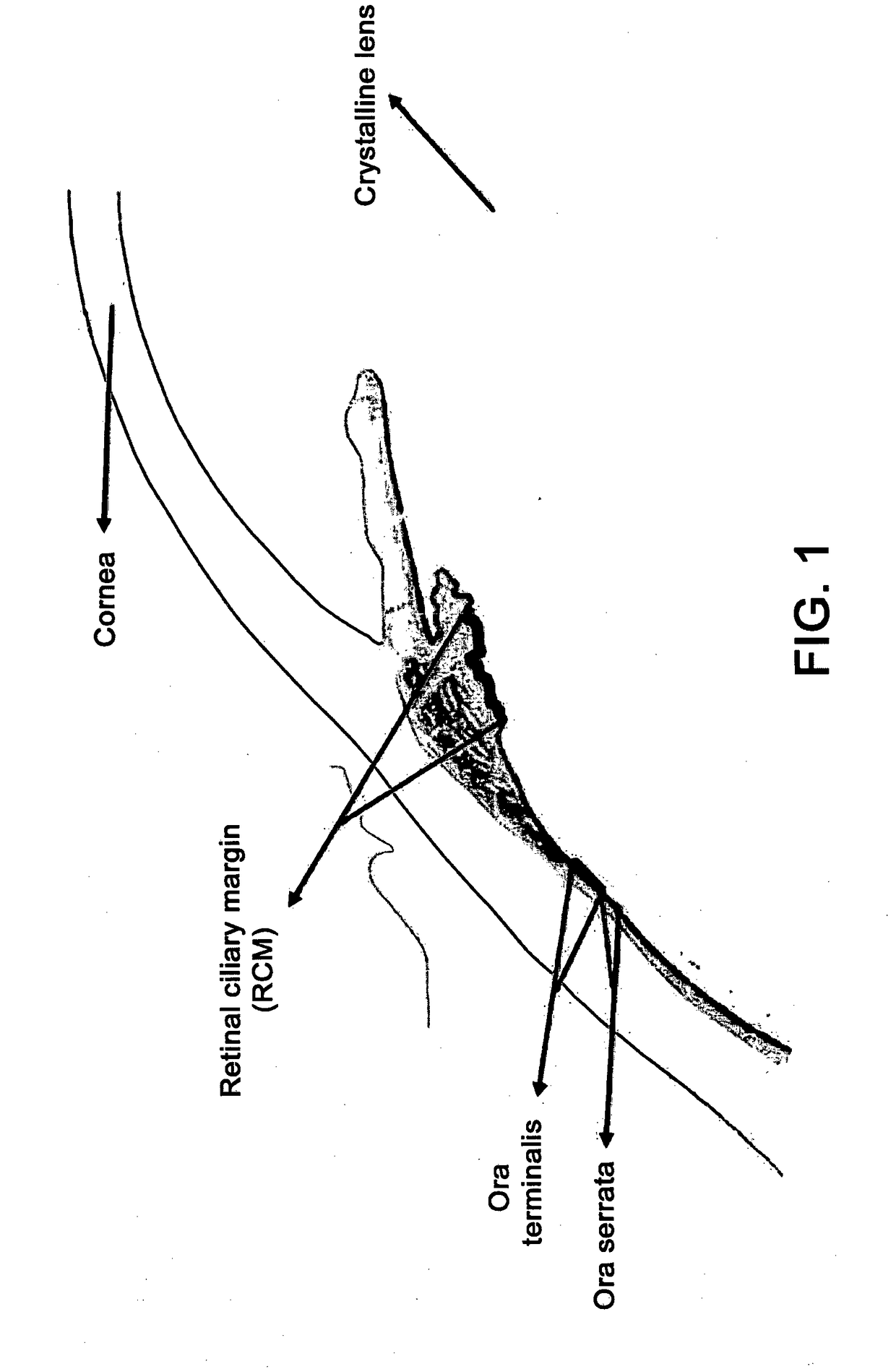
[0081]It should be noted that the stem cells are isolated exclusively from the tissue that is anatomically below the sclerocorneal limbus and practically in the ora terminalis. These cells were isolated with a frequency of approximately 1:500.
[0082]The biopsy of the tissue was then placed in a tube containing the enzymes trypsin and hyaluronidase and incubated in a water bath at 37° C. for 15 minutes. The enzymes were then blocked by means of inhibitors and single retinal stem cells were subsequently isolated with a mechanical isolation by delicate pipetting. The cells were then counted in a hemocytometric chamber, pelletized by centrifugation and resuspended in an appropriate volume of saline solution buffered with Du...
example 2
[0084]In the present experiment, in vitro proliferation, long-term self-renewal and potential differentiation of isolated ora terminalis retinal stem cells (OTRSC) were evaluated; both the sphere-forming assay and the single-layer assay were used.
[0085]The single primary cells obtained from the ora terminalis were plated on plates with 96 wells at a density of one cell per well in order to be able to test whether the isolated OTRSC were capable of proliferating in order to form clonally derived spheres. The OTRSC gave rise to clonal spheres containing both pigmented and non-pigmented cells. Using the single sphere passage assay, the primary spheres were dissociated and replanted, and the individual spheres exhibited a self-renewal capacity, each single sphere giving rise to one or more new spheres in each subsequent passage. The ora terminalis region of the human eye contains retinal stem cells that demonstrate a compliance with the in vitro growth factor that is similar to that of ...
example 3
[0088]In the present experiment, the in vivo potential of isolated OTRSC and of their progeny was evaluated.
[0089]The spherical dissociated human retinal cells containing OTRSC were transplanted into the eye of adult rabbits. In order to visualize the OTRSC and their progeny so as to transplant them into the eye of the host rabbit, a lentiviral construct was prepared which contained an enhanced green fluorescent protein (EGFP). The OTRSC were infected with the lentiviral particles which lead to the development of the green fluorescent spheres. The green fluorescent spheres were then dissociated into individual cells and resuspended in a balanced Hank saline solution at a concentration of 20,000 cells per 1 μl. Subsequently, by means of an intravitreal injection, approximately 10,000 cells per 0.5 μl were transplanted into the vitreous cavity of an eye of an adult rabbit, which had been previously anesthetized topically with proparacaine drops. These OTRSC were injected by using a pi...
PUM
 Abstract
Description
Claims
Application Information