Spatially distinguished, multiplex nucleic acid analysis of biological specimens

Active Publication Date: 2019-07-04
SPATIAL TRANSCRIPTOMICS +1
View PDF4 Cites 91 Cited by
  • Summary
  • Abstract
  • Description
  • Claims
  • Application Information

AI Technical Summary

Problems solved by technology

However, these tools are more prominent in research labs having not yet matured to the level of being readily available to the vast majority of oncologists.
Leading to even more complex mosaics within the tumor.
The end result is often an initial positive response of one subpopulation of cells, resulting in the observation of the patient's tumor shrinki

Method used


Image

  • Spatially distinguished, multiplex nucleic acid analysis of biological specimens
  • Spatially distinguished, multiplex nucleic acid analysis of biological specimens
  • Spatially distinguished, multiplex nucleic acid analysis of biological specimens

Examples


example i

Spatially Tagging mRNA from a Tissue Sample Using Illumina Flow Cells

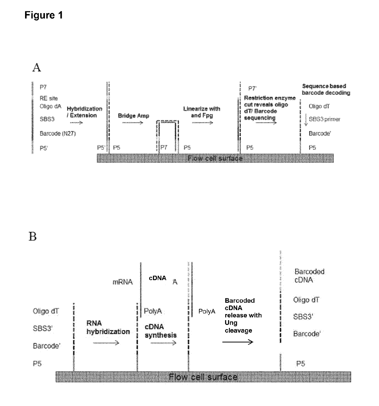
[0129]A method for generating barcoded oligo-dT containing clusters, then revealing the barcoded oligo-dT with a restriction enzyme digest followed by sequencing is described in FIG. 1. A library of fragments containing a single stranded, barcoded oligo-dA, P5′,P7, SBS3 sequencing primer binding site and a BspH1 restriction enzyme site (shown in the top panel of FIG. 1) were prepared by oligo synthesis (Integrated DNA Technologies). The barcodes were 27mers and were randomly generated during synthesis. The binding site for the SBS3 sequencing primer was included for decoding of the barcode by sequencing. An oligo-dA stretch was included to generate an oligo dT site upon clustering and linearization. Bridge amplification and clustering were performed according to standard cluster chemistry (Illumina TruSeq PE Cluster Kit v3 cBot P / N: 15037931) on an Illumina GA flow cell using manufacture's recommended protocol.

[013...

example ii

Cell Adhesion on Illumina Flow Cells

[0134]Single cells were captured on a patterned flow cell (HiSeq X10 flow cell, Illumina). All reagent flow steps were performed using a peristaltic pump or the cBOT cluster generation instrument (Illumina). Briefly, nuclease free water was flowed on all lanes of the patterned flow cell followed by 30-70K Poly D Lysine Solution (100 μg / ml and 20 μg / ml) at a flow rate of 100 μl / min for 8 min. Heat inactivated Fetal Bovine Serum (Life Technologies #10082-139) was also tested as an adhesive. The adhesives were incubated on the flow cell lanes for 1 hr, followed by a 1× PBS+0.5% Pluronic F-68 (Life Technologies #24040-032) wash. Next, the cells were adhered to the coated flow cells by flowing 5 to 50 cells / μl or approximately 100-1000 cells per lane at a rate of 100 μl / min, followed by an incubation step for 60 min to bind the cells. The flow cell was washed with 1× PBS / 0.5% pluronic at a rate of 75 μl / min. If cells were fixed on the flow cell, 1% Par...

example iv

Spatially Localized Capture of Target mRNA by Probes Attached to a BeadArray™ Surface

[0140]This example describes placement of tissue slices on top of a BeadArray™ having poly T probes, release of RNA from the tissue sections, capture of the released mRNA by the poly T probes, reverse transcription to Cy5 label the poly T probes, removal of the tissue and imaging of the BeadArray™.

[0141]As shown in Panel A of FIG. 8, a mouse olfactory tissue section was placed on a BeadArray™ having polyT probes. The tissue was treated to release mRNA and poly A tails of the released mRNA were hybridized to poly T sequences of the probes. The poly T sequences were extended using the captured mRNAs as templates and the extended primers were selectively labeled with Cy5. The tissue was removed from the BeadArray™ and the BeadArray™ was imaged to detect Cy5 flourescence.

[0142]As shown in Panel B of FIG. 7, areas of the BeadArray™ that were proximal to areas of the tissue that released mRNA species appe...


PUM

PropertyMeasurementUnit
Lengthaaaaaaaaaa
Lengthaaaaaaaaaa
Diameteraaaaaaaaaa

Abstract

A method for spatially tagging nucleic acids of a biological specimen, including steps of (a) providing a solid support comprising different nucleic acid probes that are randomly located on the solid support, wherein the different nucleic acid probes each includes a barcode sequence that differs from the barcode sequence of other randomly located probes on the solid support; (b) performing a nucleic acid detection reaction on the solid support to locate the barcode sequences on the solid support; (c) contacting a biological specimen with the solid support that has the randomly located probes; (d) hybridizing the randomly located probes to target nucleic acids from portions of the biological specimen; and (e) modifying the randomly located probes that are hybridized to the target nucleic acids, thereby producing modified probes that include the barcode sequences and a target specific modification, thereby spatially tagging the nucleic acids of the biological specimen.

Description

CROSS-REFERENCE TO RELATED APPLICATIONS[0001]The present application is a 371 of International Application No. PCT / EP2016 / 057355, filed on Apr. 6, 2016, which claims the benefit of U.S. Provisional Application No. 62 / 145,874.BACKGROUND[0002]One of every four men will die of cancer. Further statistics from the American Cancer Society predict that one of every five women will suffer the same fate. Treatments are available for many cancers. However, success for most relies on early detection.[0003]Cancer is now said to be a disease of the genome. Many oncologists and cancer researchers hope that advances in genomic analysis tools will provide early detection and a path to treatment. However, these tools are more prominent in research labs having not yet matured to the level of being readily available to the vast majority of oncologists. Improvements are needed.[0004]It has been said that at the time of diagnosis, all cancer patients are mosaics. They are mosaics because they have at le...

Claims


Application Information

Patent Timeline
IPC IPC(8): C12Q1/6834C12Q1/6841C12Q1/6876C12Q1/6874
CPCC12Q1/6834C12Q1/6841C12Q1/6876C12Q1/6874C12Q2525/179C12Q2563/149C12Q2525/173C12Q2543/101C12Q2565/515C12Q2525/131C12Q2535/122C12N15/1065C12Q2543/10
Inventor FRISEN, JONASST HL, PATRIKLUNDEBERG, JOAKIMCANN, GORDON M.BAZARGAN, LEILAARAVANIS, ALEX
Owner SPATIAL TRANSCRIPTOMICS
Why Eureka
Try Eureka