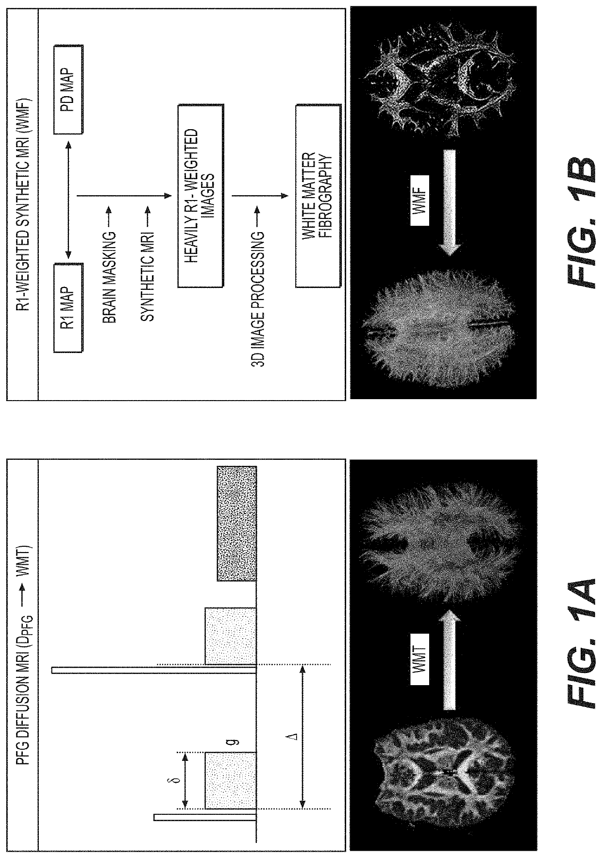
White matter fibrography by synthetic magnetic resonance imaging
a technology of synthetic magnetic resonance imaging and white matter, applied in the field of white matter fibrography by synthetic magnetic resonance imaging, can solve the problems that r1-weighted pulse sequences may be difficult to implement in actual (physical) physical environments, and achieve the effect of enhancing the subtle wm textur
- Summary
- Abstract
- Description
- Claims
- Application Information
AI Technical Summary
Benefits of technology
Problems solved by technology
Method used
Image
Examples
example 1
Platform Independence
[0092]As a first step for validating WMF, Mill scanning platform independency was shown by analyzing imaging data at a medium-high spatial resolution (voxel=0.5×0.5×2 mm3). Comparable quality imaging data generated with two 3.0 T Mill scanners of different manufacturers was processed in under 8 min of scan time each. The resulting connectome renditions of the two healthy volunteers shown in FIG. 15 have comparable quality in terms of signal-to-noise, fiber delineation, left-right symmetry, and organization. Also noticeable is the high fiber density in the prefrontal lobe, which characterizes the adult human brain.
example 2
f Advancing Age (1.5 T)
[0093]A second WMF validation step illustrates the normal brain age-related changes: whole brain axial connectome renditions as a function of increasing age are shown in FIG. 16 using lower spatial resolution (voxel=0.94×0.94×3 mm3) data generated at 1.5 T. High organizational level and basic bilateral symmetry is observed at all ages. Myelination progresses at a very fast pace during the first two years of life during infancy to toddler's first year. The genu of the corpus callosum (CC) is clearly visible at 0.6 years and myelinates anteriorly-to-posteriorly with the CC splenium becoming clearly discernable as early as 1.3 years of age. Globally, the posterior aspect of the brain is markedly more myelinated during infancy and myelination progresses in the posterior-to-anterior direction as well as from the brain's center to the periphery towards the cortex; WM fibers become thinner as these approach the cortex. From early adolescence to young adulthood, the p...
example 3
Stroke (3.0 T)
[0094]A third WMF technique validation step assesses WM integrity under the stress of ischemia. Two acute ischemic stroke lesions (arrow) in a 48 yo female are shown in FIG. 17 via standard dMRI as bright lesions in diffusion weighted image (DWI) and dark lesions in the map of the apparent diffusion coefficient (ADC) thus confirming restricted diffusion. The two lesions appear as signal voids in the R1-weighted image likely indicating irreversible WM obliteration at this stage 13 hours after last time seeing well.
PUM
 Abstract
Description
Claims
Application Information
 - R&D
- Intellectual Property
- Life Sciences
- Materials
- Tech Scout
- Unparalleled Data Quality
- Higher Quality Content
- 60% Fewer Hallucinations
Browse by: Latest US Patents, China's latest patents, Technical Efficacy Thesaurus, Application Domain, Technology Topic, Popular Technical Reports.
© 2025 PatSnap. All rights reserved.Legal|Privacy policy|Modern Slavery Act Transparency Statement|Sitemap|About US| Contact US: help@patsnap.com


