Localization and characterization of subsurface structures using temporally-resolved photon density waves

a technology of photon density wave and subsurface structure, applied in medical science, surgery, diagnostics, etc., can solve the problems of difficult ultrasound technology for precise tracking and localization of thin needles buried in turbid tissues, difficult procedure, and persistent challenge of needles on ultrasound, so as to improve sensitivity and information content, the effect of greater precision and more safe operation

Inactive Publication Date: 2020-01-09
RGT UNIV OF CALIFORNIA
View PDF0 Cites 0 Cited by
  • Summary
  • Abstract
  • Description
  • Claims
  • Application Information

AI Technical Summary

Benefits of technology

[0012]According to some embodiments, single or multiple frequencies and single or multiple wavelengths may be utilized to improve sensitivity and information content. One or more photo detectors at the surface can be used to solve for the position of the needle in N-dimensional space. Once the position of the needle is known, the data can be integrated with ultrasound data, providing real-time guidance of the needle or catheter to an ultrasound identified target. This may be helpful when determining the location of the needle is critical to the success of a procedure. For example, this method may be used to help guide clinicians to particular targets inside tissue, such as for delivering anesthetic drug to a specific nerve site or guiding the needle to a biopsy site. This method has specific advantages when the needle is difficult to visualize and track using ultrasound. In addition, this method for delivery of light deep in tissue to a subsurface object or structure can be used to characterize specific features of that buried object or target tissue such as the object / structure's optical absorption, scattering, and physiological properties.
[0013]One of the unique and inventive technical features of the present invention is the use of frequency domain photon migration (FDPM) methods to extract a position of the needle, allowing a procedure to be performed with greater precision. Without wishing to limit the invention to any theory or mechanism, it is believed that the technical feature of the present invention advantageously provides for sub-millimeter resolution tracking of a needle in turbid tissue. This technique may also allow a procedure (e.g. injection of drug into specific tissue sites) to be performed more safely, as it eliminates reliance on “eye-balling” by providing quantitative positional information of the needle in biological tissue. Finally, the present technique allows for characterization of the optical and physiological properties of the object, which may change upon administration of therapy via the needle and could be used as feedback for therapeutic guidance and dosing. By using amplitude and phase information acquired by FDPM, this technique will provide sub-millimeter resolution tracking of a needle in turbid tissue. Due to the nature of near-infrared light, this method is appropriate for use in turbid tissue up to several centimeters deep. Trilateration of the needle is also possible, and can be integrated with ultrasound, allowing physicians to simultaneously access physiological information, as well as the position of the needle in the ultrasound field of view. None of the presently known prior references or work has the unique inventive technical feature of the present invention.

Problems solved by technology

Precise tracking and localization of thin needles buried in turbid tissues is difficult using ultrasound technology due to lack of contrast.
However, visualizing the needle on the ultrasound is a persistent challenge and complicates the procedure.
This previously explored technique utilized line-of-sight detection (i.e. human vision) and intensity information to localize the needle, thus quantitative positional information is unavailable.
Attenuation of the light can be the result of many factors including needle depth and tissue optical properties, making it difficult to accurately track the needle location in thick turbid tissues and thereby limiting the Cha technique to applications involving thin, homogeneous, or relatively transparent tissues.

Method used


Image

  • Localization and characterization of subsurface structures using temporally-resolved photon density waves
  • Localization and characterization of subsurface structures using temporally-resolved photon density waves
  • Localization and characterization of subsurface structures using temporally-resolved photon density waves

Examples


example

[0042]The following is a non-limiting example of an optical method to track the position of a needle catheter inside of tissue. It is to be understood that said example is not intended to limit the invention in any way, and that equivalents or substitutes are within the scope of the invention.

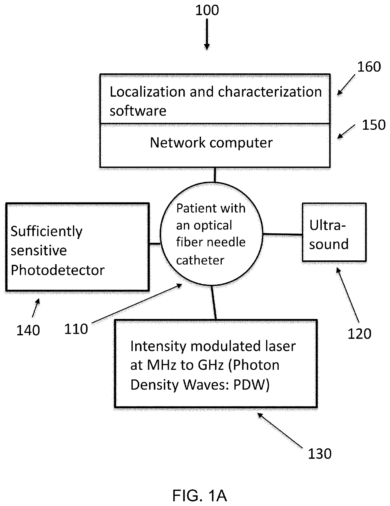
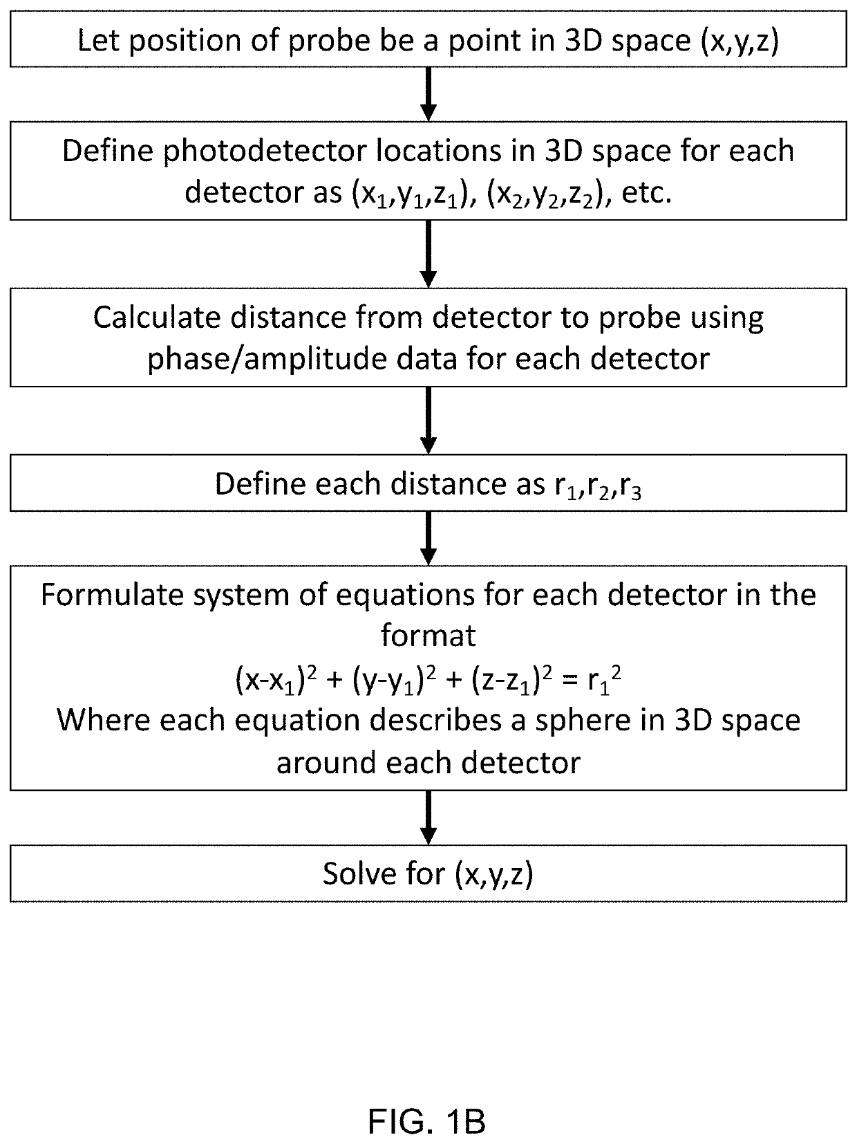
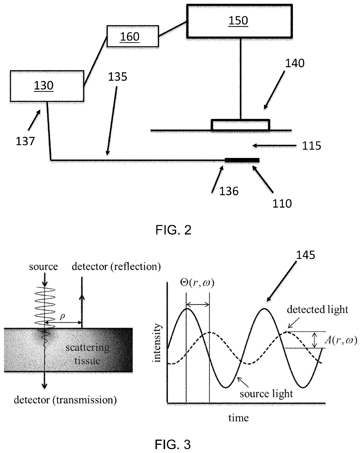
[0043]Frequency domain photon migration (FDPM) is an optical technique that illuminates tissue with high-frequency (e.g. 50 MHz to 600 MHz) intensity-modulated near-infrared lasers and detects changes in the light scattered back from the tissue to determine tissue optical properties. When frequency domain detection is utilized, the amplitude (A) and phase shift (φ) of the detected light can be used to determine the position of a light source delivered inside the needle tip using a thin optical fiber. This is due to the fact that the phase shift (φ) of the intensity modulated light is linearly proportional to the distance (d) between the light source (i.e. the needle tip) and the detector, where...


PUM


Abstract

Optical systems and methods to track the position of a needle in subsurface structures, such as tissues or organs, and co-register the information with ultrasound are described herein. An optical fiber in a needle catheter is used to transmit light inside of the structure. The light is intensity modulated at sufficiently high frequencies such that the time of arrival of the light can be used to determine the distance of the needle from an optical detector at the tissue surface. The position of the needle can be tracked by combining data obtained using different modulation frequencies and/or wavelengths of light. By using multiple detectors at different positions, the location of the needle in 3D space can be triangulated using light, and the data can be integrated with ultrasound to obtain the anatomical structure.

Description

CROSS-REFERENCES TO RELATED APPLICATIONS[0001]This application is a non-provisional, and claims benefit of U.S. Provisional Patent Application No. 62 / 694,689, filed Jul. 6, 2018, the specification of which is incorporated herein in its entirety by reference.GOVERNMENT SUPPORT[0002]This invention was made with Government support under Grant No. P41EB015890, awarded by the National Institutes of Health and Grant No. FA9550-17-0193, awarded by the U.S. Air Force Office of Scientific Research. The Government has certain rights in the invention.FIELD OF THE INVENTION[0003]The present invention relates to systems and methods for tracking and localizing subsurface tissue probes, such as needles, and also characterizing subsurface objects, such as tumors, using temporally-resolved diffuse photon density waves.BACKGROUND OF THE INVENTION[0004]Precise tracking and localization of thin needles buried in turbid tissues is difficult using ultrasound technology due to lack of contrast. In a typic...

Claims


Application Information

Patent Timeline
Patent Type & Authority Applications(United States)
IPC IPC(8): A61B17/34A61B8/00
CPCA61B17/3403A61B8/466A61B2017/3413A61B8/12A61B2090/373A61B2090/3735A61B2090/3945
Inventor TROMBERG, BRUCE J.LAM, JESSEQUANG, TIMOTHY
Owner RGT UNIV OF CALIFORNIA
Why Eureka
Try Eureka