
Computer-aided diagnostic system for early diagnosis of prostate cancer

a prostate cancer and diagnostic system technology, applied in computing, genomics, instruments, etc., can solve the problems of high dre cost, and inability to detect prostate cancer through dr

Active Publication Date: 2020-01-09
UNIV OF LOUISVILLE RES FOUND INC
View PDF0 Cites 10 Cited by
  • Summary
  • Abstract
  • Description
  • Claims
  • Application Information

AI Technical Summary

Benefits of technology

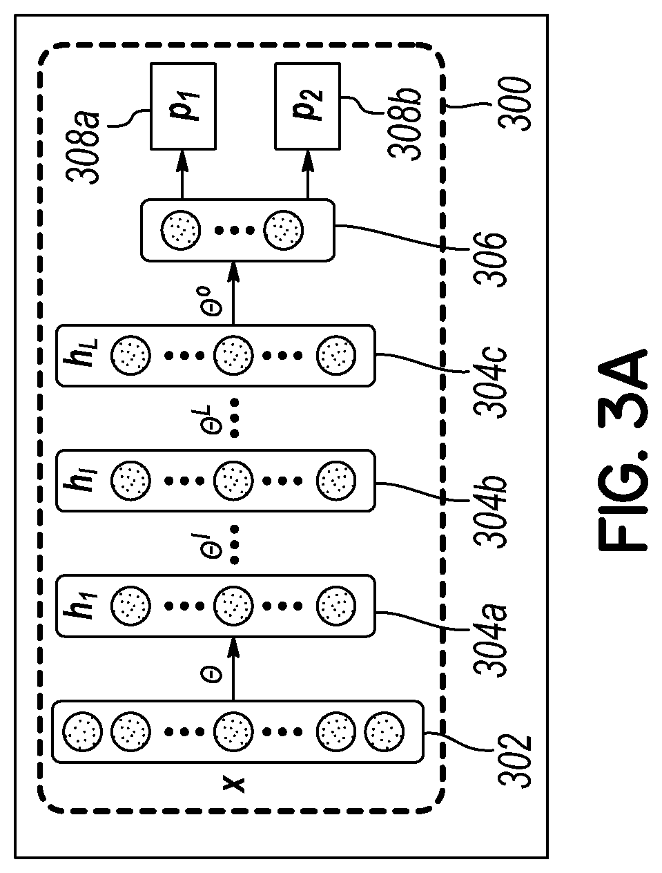
The present invention provides a method and system for diagnosing prostate cancer using magnetic resonance imaging prostate data and biological output probability data. The system obtains the magnetic resonance imaging prostate data at multiple b-values and processes it through multiple autoencoders to create a output probability data. The output probability data is then organized into an input vector and processed through an autoencoder to generate a diagnosis of prostate cancer. The technical effect of the invention is an improved diagnosis accuracy and sensitivity for prostate cancer using magnetic resonance imaging and biological data.

Problems solved by technology

The cost of DRE is relatively low, but DRE is highly invasive.
However, most of the central-zone and transitional-zone tumors, as well as tumors too small to be palpated, cannot be detected through DRE.
However, both DRE and PSA are merely indicators of prostate cancer and are not conclusive diagnosis techniques.
However, there is a possibility of missing a cancer tumor due to the small number of biopsy specimens, random nature of sampling, and poor resolution of TRUS.
Even though biopsy is the most precise technique for detecting cancer, it is highly invasive, expensive, and a painful tool for detecting prostate cancer and determining its aggressiveness.
Therefore, it is hard to precisely detect tumors and / or identify the cancer stage using TRUS images.
There are two main limitations of the developed CAD systems for prostate cancer diagnosis from DW-MRI.
First, most of these CAD systems only use images acquired at a certain b-value.
Therefore, there is no agreement on which b-value will provide the best diagnosis.
Second, most investigators just use imaging markers and do not integrate them with the current clinical biomarkers, which may affect the final accuracy of the diagnosis.

Method used


Image

  • Computer-aided diagnostic system for early diagnosis of prostate cancer
  • Computer-aided diagnostic system for early diagnosis of prostate cancer
  • Computer-aided diagnostic system for early diagnosis of prostate cancer

Examples


Embodiment Construction

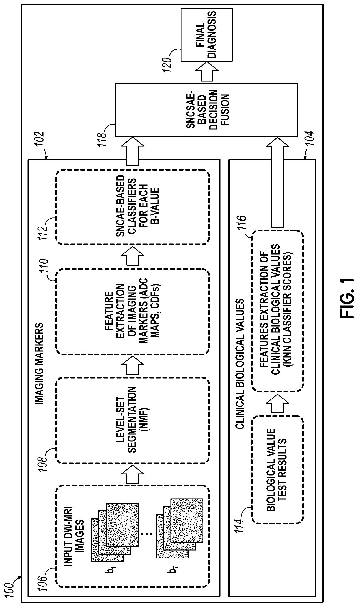
[0030]Embodiments of the invention comprise methods, systems, and computer program products for analyzing medical images (e.g., prostate image scans) of a medical imaging scan and analyzing biological values (e.g., prostate specific antigen levels) of a clinical biological test. The limitations of existing diagnostic methods are addressed by integrating imaging markers with clinical biomarkers to provide an accurate and robust system for early diagnosis of prostate cancer. DW-MRI data collected at multiple b-values may be used to reduce sensitivity to the selection of a b-value. A deep learning technique may be used to fuse images acquired at multiple b-values with clinical biomarkers to provide a diagnosis of prostate cancer.

[0031]In some embodiments of the invention, medical images for a magnetic resonance image (MRI) scan of a prostate may be analyzed, and a probable diagnosis of cancer may be specified. In other embodiments, the probable diagnosis of cancer based on the MRI scan...


PUM


Abstract

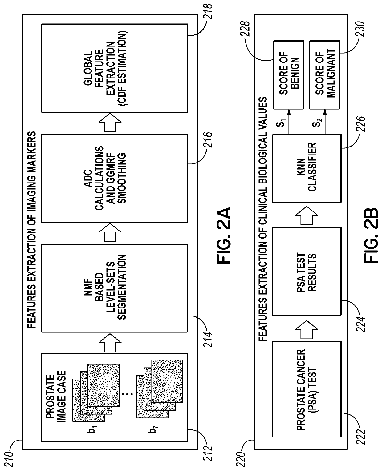
Systems and methods for diagnosing prostate cancer. Image sets (e.g., MRI collected at one or more b-values) and biological values (e.g., prostate specific antigen (PSA)) have features extracted and integrated to produce a diagnosis of prostate cancer. The image sets are analyzed primarily in three steps: (1) segmentation, (2) feature extraction, smoothing, and normalization, and (3) classification. The biological values are analyzed primarily in two steps: (1) feature extraction and (2) classification. Each analysis results in diagnostic probabilities, which are then combined to pass through an additional classification stage. The end result is a more accurate diagnosis of prostate cancer.

Description

CROSS-REFERENCE TO RELATED APPLICATIONS[0001]This application claims the benefit of and priority to U.S. Provisional Application No. 62 / 529,772 entitled COMPUTER-AIDED DIAGNOSTIC SYSTEM FOR EARLY DIAGNOSIS OF PROSTATE CANCER, filed Jul. 7, 2017, which is expressly incorporated by reference herein in its entirety.FIELD OF THE INVENTION[0002]This disclosure generally relates to computer analysis of medical image data and clinical data, and in particular to the analysis of medical images of the prostate and biological values that combined lead to a diagnosis related to prostate cancer based.BACKGROUND OF THE INVENTION[0003]Prostate cancer is one of the most frequent cancers identified among the male population in the United States, and accounts for the highest mortality rate due to cancer second only to lung cancer. According to statistics published by the American Cancer Society in 2016, around 180,890 new patients were diagnosed and around 26,120 patients passed away due to prostate ...

Claims


Application Information

Patent Timeline
Patent Type & Authority Applications(United States)
IPC IPC(8): G06F19/24G06F19/12
CPCG16B5/00G16B40/00G16B20/00G16B40/20G16B40/30
Inventor EL-BAZ, AYMAN S.SHALABY, AHMEDKHALIFA, FAHMIABDELMAKSOUD, ISLAM
Owner UNIV OF LOUISVILLE RES FOUND INC
Learn More