Eureka AIR delivers breakthrough ideas for toughest innovation challenges, trusted by R&D personnel around the world.

Detection and/or correction of residual iodine artifacts in spectral computed tomography (CT) imaging

Active Publication Date: 2020-01-23
KONINKLJIJKE PHILIPS NV
View PDF0 Cites 0 Cited by
  • Summary
  • Abstract
  • Description
  • Claims
  • Application Information

AI Technical Summary

Benefits of technology

The invention described in this patent addresses certain problems and provides a solution.

Problems solved by technology

However, with conventional CT, the projection data does not reflect the spectral characteristics as the data it represents is proportional to the energy fluence integrated over the energy spectrum.
However, VNC images display isolated residual iodine artifacts, particularly in the region of small vessels in the liver, when generated with spectral components decomposed from de-noised spectral image data.
Unfortunately, these artifacts reduce the confidence of the clinician regarding the accuracy of the VNC images in other areas.

Method used


Image

  • Detection and/or correction of residual iodine artifacts in spectral computed tomography (CT) imaging
  • Detection and/or correction of residual iodine artifacts in spectral computed tomography (CT) imaging
  • Detection and/or correction of residual iodine artifacts in spectral computed tomography (CT) imaging

Examples


Embodiment Construction

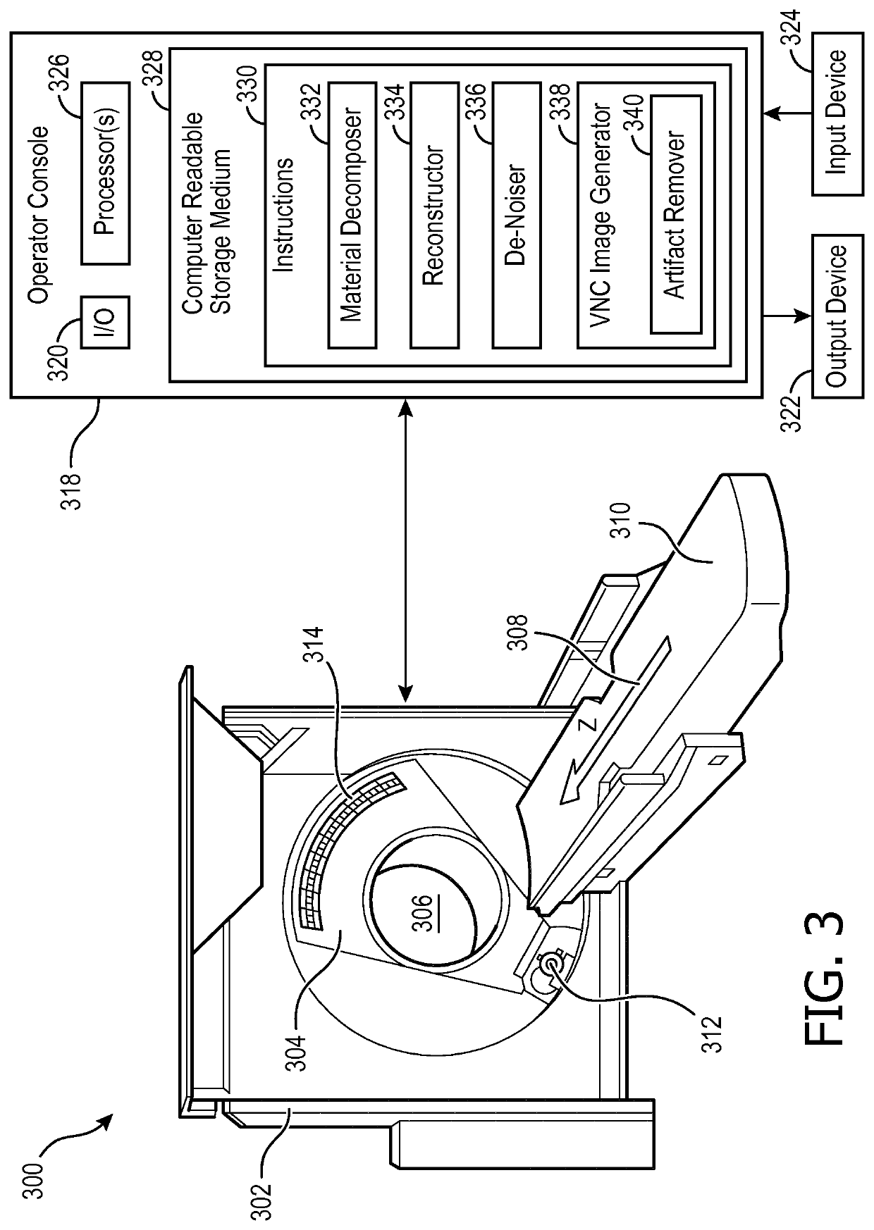
[0035]FIG. 3 schematically illustrates an imaging system 300 such as a computed tomography (CT) scanner configured for spectral (multi-energy) imaging.

[0036]The imaging system 300 includes a stationary gantry 302 and a rotating gantry 304. The rotating gantry 304 is rotatably supported by the stationary gantry 302 and rotates around an examination region 306 about a longitudinal or z-axis 308. A subject support 310, such as a couch, supports an object or subject in the examination region. The subject support 310 is movable in coordination with performing an imaging procedure so as to guide the subject or object with respect to the examination region 306. The following is described in the context of a contrast-agent (e.g., iodine) scan of an object or a subject administered the contrast agent.

[0037]A radiation source 312, such as an x-ray tube, is rotatably supported by the rotating gantry 304. The radiation source 312 rotates with the rotating gantry 304 and emits poly-chromatic rad...


PUM


Abstract

A system (300) includes input / output configured to receive line integrals from a contrast enhanced spectral scan by an imaging system. The system further includes (300) a processor (326) configured to: decompose (334) the line integrals into at least Compton scatter and a photo-electric effect line integrals; reconstruct the Compton scatter and a photo-electric effect line integrals to generate spectral image data, including at least Compton scatter and photo-electric effect images; de-noise (332) the Compton scatter and photo-electric effect images; identify (402) residual iodine voxels in the de-noised Compton scatter and the photo-electric effect images corresponding to residual iodine artifact; and produce a virtual non-contrast image using the identified residual iodine voxels

Description

FIELD OF THE INVENTION[0001]The following generally relates to spectral imaging and more particularly to detection and / or correction of residual iodine artifacts in spectral computed tomography (CT) imaging.BACKGROUND OF THE INVENTION[0002]A CT scanner generally includes an x-ray tube mounted on a rotatable gantry opposite one or more detectors. The x-ray tube rotates around an examination region located between the x-ray tube and the one or more detectors and emits radiation that traverses the examination region and a subject and / or object disposed in the examination region. The one or more detectors detect radiation that traverses the examination region and generate a signal or projection data indicative of the examination region and the subject and / or object disposed therein.[0003]The projection data is reconstructed to generate volumetric image data, which can be used to generate one or more images. The resulting image(s) includes pixels that are represented in terms of gray sca...

Claims


Application Information

Patent Timeline
IPC IPC(8): G06T11/00G06T5/50G06T5/00
CPCG06T11/005G06T11/008G06T2207/20192G06T2207/20216G06T2207/20224G06T5/50G06T2207/30008G06T2207/10081G06T2211/424G06T5/002G06T11/006G06T2211/408G06T5/70
Inventor BROWN, KEVIN MARTINBRENDEL, BERNHARD JOHANNES
Owner KONINKLJIJKE PHILIPS NV
Why Eureka
Eureka Blog
Learn More