
Cell culture device and methods

Pending Publication Date: 2020-02-27
MIMETAS BV
View PDF1 Cites 5 Cited by
  • Summary
  • Abstract
  • Description
  • Claims
  • Application Information

AI Technical Summary

Benefits of technology

The patent text is explaining a method for controlling the movement of droplets in a system. By using a capillary pressure barrier, the droplets can be confined to a specific location. The technical effect of this method is that it allows for better control and manipulation of droplets, which can be useful in various applications such as chemical synthesis and biological research.

Problems solved by technology

Although it is claimed that these spheroids have significantly improved predictivity to standard cell cultures, it is not used for most organoid cultures.
As already addressed above these organoids lack the presence of vasculature, thus limiting their growth as beyond a certain size hypoxic and in a later stage necrotic cores may be formed.
None of the above examples of microfluidic cell culture allows for culture of organoids in an ECM with distinct vasculature that can be perfused.
The progress of growing organoids and embryonic bodies, is hampered by lack of technical means to support this growth.
The culture techniques are highly cumbersome and may vary greatly from organ to organ.
Third, the extracellular matrix (ECM) can be a crucial factor for correct differentiation, but culture protocols are not all compatible with ECM gel culture.
Most importantly of all, so far the growth of embryonic bodies and organoids has been strongly limited by lack of vasculature.

Method used


Image

  • Cell culture device and methods
  • Cell culture device and methods
  • Cell culture device and methods

Examples


Embodiment Construction

[0214]The present invention will now be described by way of example only with reference to the drawings.

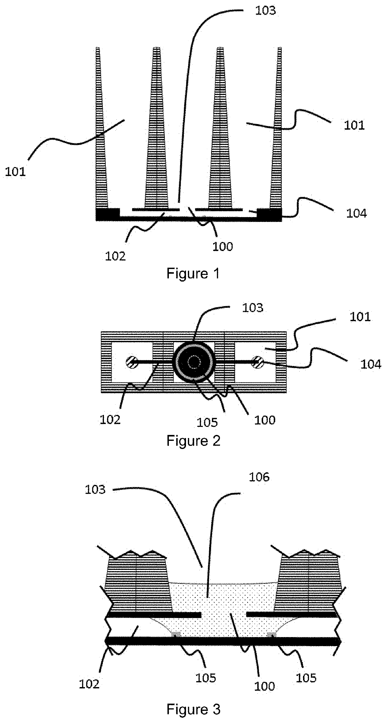
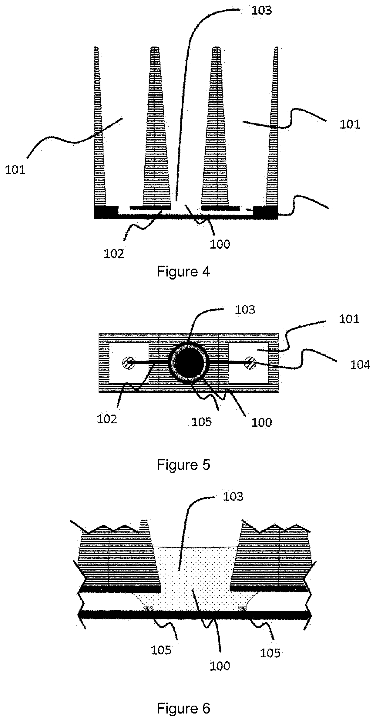
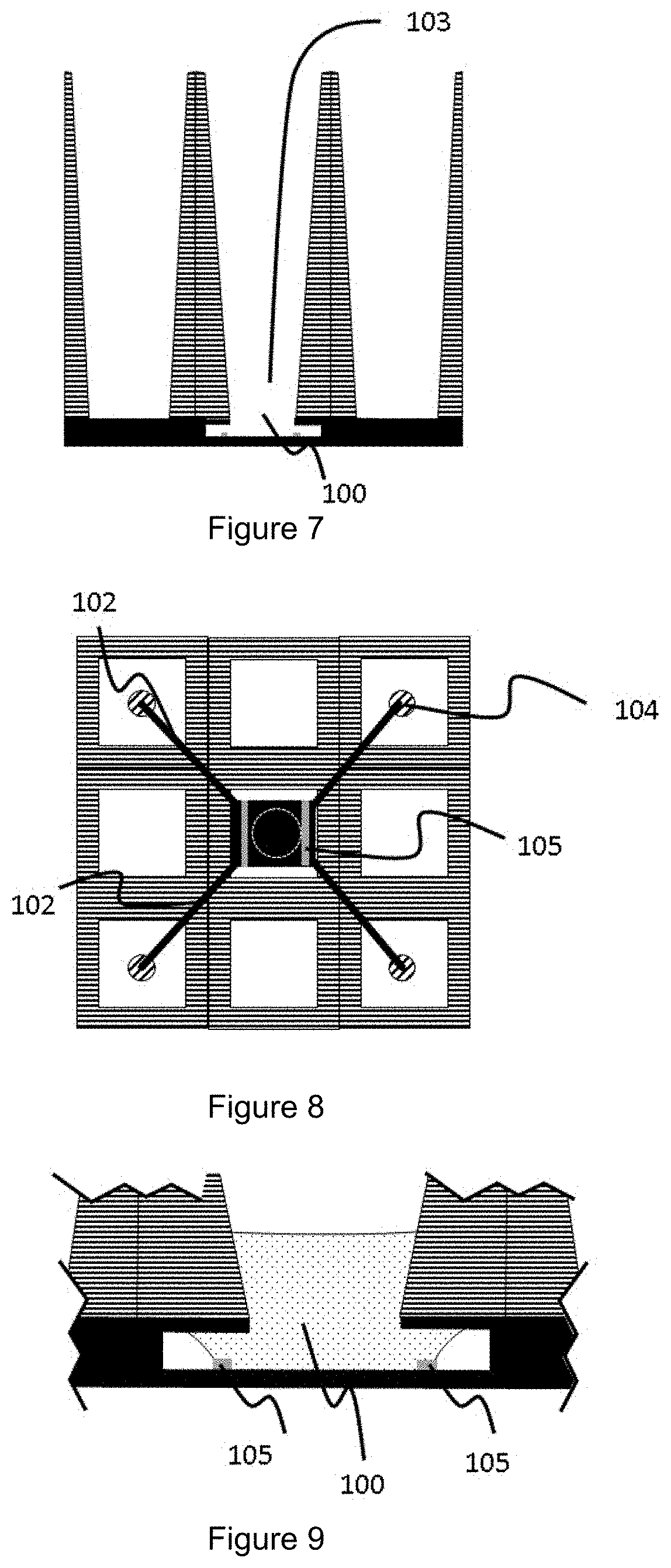
[0215]A first example of a cell culture device is schematically shown in FIGS. 1 to 3. The view across the device (FIG. 1) shows a central organoid compartment (103) flanked by two reservoirs (101) and a microfluidic channel (102) in a microfluidic layer (shown in solid) connecting the reservoirs with the organoid compartment via inlets (104) in the cover layer of the microfluidic layer. A capillary pressure barrier (105) is present at the base of the organoid compartment and accessible via aperture (100) in the cover layer of the microfluidic layer (shown in solid). In this particular example, the aperture is of narrower cross-section than the downwardly extending walls of the organoid compartment.

[0216]When charged with sufficient volume of the first liquid (the gel or gel precursor (106) (comprising one or more types of cells or cell aggregates, also termed the organoid composi...


PUM


Abstract

A method of vascularising a cell aggregate on a microfluidic device, microfluidic cell culture devices comprising perfusable vascular networks and kits and assays using the microfluidic cell culture devices are described. The microfluidic devices comprise one or more capillary pressure barriers allowing for formation of an extracellular matrix gel within a confined area of the network, in which cells can be cultured for different uses.

Description

FIELD OF THE INVENTION[0001]The present invention relates to a method of a device for 3D culture of cells, for example a multi-well plate allowing for a controlled and reliable vascularization and / or perfusion of organoid assays and / or cell cultures. It equally provides methods for generation of 3D cultured cells and for vascularization / perfusion of the cultured cells, and assay plates and uses thereof resulting from the device and methods.BACKGROUND TO THE INVENTION[0002]In a drive towards ever better predictive phenotypic models for drug testing and disease mimicking, progress has been made in recent years in the field of organoid culture. An organoid is a three-dimensional organ-bud that typically comprises most specialized cells that are also available in the human body. In practice, the culture and differentiation of tissue during embryonic regenerative development is mimicked in an in vitro environment, such that stem cells differentiate to various differentiated cells.[0003]A...

Claims


Application Information

Patent Timeline
IPC IPC(8): C12M3/06B01L3/00C12M3/00C12N5/071C12M1/00
CPCC12M21/08C12N5/0691B01L2300/041C12N2501/999C12N2506/28C12M23/16B01L3/502715B01L2300/0858C12M23/38C12N2533/30C12N2531/00C12M23/12B01L3/5027
Inventor VULTO, PAULTRIETSCH, SEBASTIAAN JOHANNESNICOLAS, ARNAUD
Owner MIMETAS BV
Learn More