System and Method for Wirelessly Transmitting Operational Data From an Endoscope to a Remote Device

a technology of wireless transmission and endoscope, which is applied in the field of medical devices, can solve the problems of limiting the use of the endoscope to procedures conducted, unable to capture images and videos, and extremely limited mobility, and achieves the effect of small footprint and not cost effectiv

Inactive Publication Date: 2020-06-18
INTEGRATED MEDICAL SYST INT INC
View PDF0 Cites 3 Cited by
  • Summary
  • Abstract
  • Description
  • Claims
  • Application Information

AI Technical Summary

Benefits of technology

[0014]One advantage of the present invention is its adaptability. For example, wireless transmission of operational data allows an examiner to monitor an ongoing operation using the examiner's personal device, such as: a smart phone, a tablet, a head-mounted display, or a monitor.
[0015]Remote monitoring of an endoscopy procedure provides yet another advantage of the many embodiments by enabling classrooms or seminars to participate in a live operation. This opens up new possibilities where only a passive review of prerecorded operations was previously possible. Clinical studies may be expanded beyond centralized operational facilities to remote sites, such as a battlefield, emergency clinic, or even a barn. When coupled with the operational data sharing method discussed in detail below, the remote networking capabilities enable new and useful telemedicine applications. For example, an experienced physician could oversee multiple concurrent off-site operations conducted by junior physicians, and provide operational feedback through his monitoring device.
[0016]Another advantage of the present invention is its portability. For human patients, endoscopy procedures are performed in centralized facilities, such as a hospital or clinic, where the equipment may be stored and operated. It is reasonable to expect a patient to travel to and from the facility to have the operation performed. However, for veterinarians performing similar operations, it is not cost effective to transport a large animal, such as a horse or a cow, to a clinic or animal hospital. This is especially true for large marine animals, such as a whale or dolphin. Accordingly, the relatively small footprint of the many embodiments enables veterinarians to travel off-site to perform endoscopy operations. Furthermore, it enables veterinarians to schedule the examination of multiple animals at the same site, or schedule multiple operations in the same day and travel from site to site.
[0017]In order to achieve the main objective, the present invention is directed to the satisfaction of the capabilities required from a conventional endoscope comprising a main body, an insertion tube, valves connected to guide channels to support air, water, and therapeutic instruments, therapeutic instrument insertion port, and angulation knobs and componentry; all of which comprise an existing FDA approved medical device.

Problems solved by technology

Such a configuration limits the usage of the endoscope to procedures conducted in the examiner's office due to the size and weight of the supporting equipment.
Mobility is extremely limited due to its bulky nature.
Additionally, this option, by itself, does not provide the capability to capture images and video, nor does it allow for printing or the possibility of other integrated functionality.
Use with an eyepiece can also present unique challenges for veterinarians, who must stand in close proximity to an animal patient, which may become spooked during the operation.
Such an encapsulated approach leads to expensive proprietary solutions that handcuff the display technology to the signal processing unit and preclude the substitution of other general purpose displays such as a smart device or tablet.

Method used


Image

  • System and Method for Wirelessly Transmitting Operational Data From an Endoscope to a Remote Device
  • System and Method for Wirelessly Transmitting Operational Data From an Endoscope to a Remote Device
  • System and Method for Wirelessly Transmitting Operational Data From an Endoscope to a Remote Device

Examples


Embodiment Construction

[0038]Referring to the drawings, embodiments of the present invention will be described below.

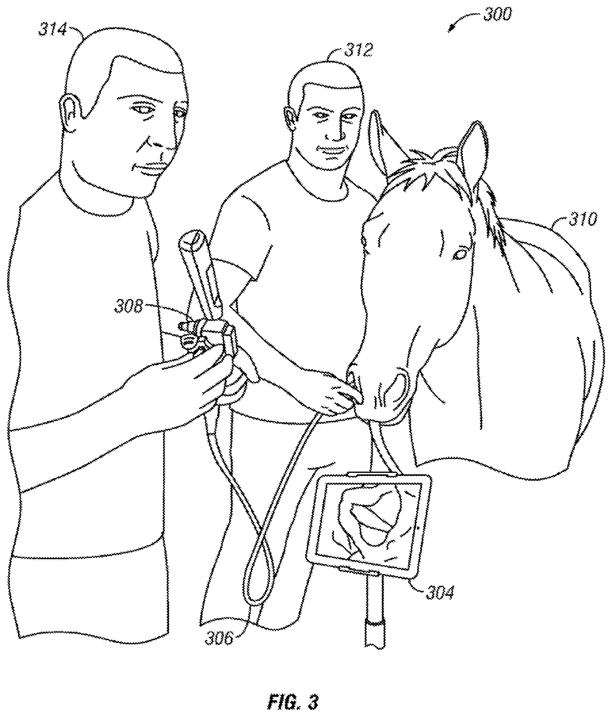
[0039]FIG. 3 illustrates a respiratory endoscopy examination being performed on a horse, indicated generally at 300, using a wireless endoscope 308 connected to a mobile device 304 in accordance with one embodiment. As FIG. 3 demonstrates, wireless endoscopy is particularly useful in applications where a patient is not fully sedated or restrained. During an operation, the insertion tube 306 of the wireless endoscope 308 is introduced into the horse's 310 respiratory system via its nostril. Attendant 312 stabilizes the horse 310 and guides the insertion tube 306 during the operation. The veterinarian 314 observes the operation on the display 304, which may be supported by a tripod or stand, while controlling the endoscope 308.

[0040]If a veterinary patient, such as a horse, becomes spooked during an operation, the absence of wires can reduce trauma to the animal, which has less equipment atta...


PUM


Abstract

A system for wirelessly transmitting data from an endoscope, comprising an endoscope having a control body, an insertion tube extending from the control body, the distal end of the insertion tube containing an image sensor and a light source, and a control head connected to the control body, which comprises a battery; a light source amplifier connected to the battery, the light source amplifier operable to boost the intensity of the light source; a video processor configured to create compressed video data from a video stream captured via the image sensor; and a wireless communication module configured to negotiate a wireless connection with a mobile device, wherein the wireless communication module is further configured to transmit the compressed video data to the mobile device over the wireless connection, and wherein the wireless communication module comprises a channel discriminator configured to automatically avoid RF interference.

Description

CROSS-REFERENCE TO RELATED APPLICATIONS[0001]This disclosure is a divisional of U.S. application Ser. No. 14 / 508,265, filed Oct. 7, 2014, which claims the benefit of U.S. Provisional Application No. 61 / 998,690, filed Jul. 7, 2014.BACKGROUND OF THE INVENTION1. Field of the Invention[0002]This invention relates in general to the field of medical devices, and more particularly to a system and method for wirelessly transmitting operational data from an endoscope to a remote device.[0003]Doctors and veterinarians rely on a bevy of medical imaging technologies, including x-rays, x-ray fluoroscopy, magnetic resonance imaging (MRI), and CT / PET scans to obtain different views of a patient's symptoms and anatomy. However, for some conditions, it may be advantageous or necessary to gather real-time operational data from inside the body by relying on a device known as an endoscope.[0004]Endoscopes can be used in a variety of medical procedures. For example, an endoscope may be used to investiga...

Claims


Application Information

Patent Timeline
Patent Type & Authority Applications(United States)
IPC IPC(8): A61B1/00A61B1/06A61B1/05
CPCA61B1/00016A61B1/00009A61B1/00108A61B1/05A61B1/00032A61B1/00039A61B1/0676A61B1/00048A61B1/00042
Inventor WILLIAMS, DAWN R.
Owner INTEGRATED MEDICAL SYST INT INC
Why Eureka
Try Eureka