Fat droplets in retina and optic nerve as a diagnostic marker for neurodegeneration and glaucoma in humans

a technology of retinal and optic nerve, which is applied in the field of fat droplets in retina and optic nerve as a diagnostic marker for neurodegeneration and glaucoma in humans, can solve the problems of inability to distinguish these two groups of patients, lack of reliable predictors of iop, and limited diagnostic value of methods, so as to avoid unnecessary medication and improve focus

Active Publication Date: 2020-06-25
JACKSON LAB THE
View PDF0 Cites 1 Cited by
  • Summary
  • Abstract
  • Description
  • Claims
  • Application Information

AI Technical Summary

Benefits of technology

[0010]It is an object of the present invention to provide a method for diagnosing glaucoma in a patient which comprises the steps: (a) providing a patient in needs of diagnosing glaucoma; (b) detecting the presence of fat droplets in the retina and / or optic nerve of the patient, wherein the presence of the fat droplets is predictive of the occurrence of glaucoma (diagnostic of glaucoma). The method of the invention allows better focus on caring for those who will develop glaucoma, while avoiding unnecessary medication of those who will not.

Problems solved by technology

However, elevated IOP is not a reliable predictor for glaucoma.
Currently, there is not way to distinguish these two groups of patients before there is evident visual system disease.
Furthermore, glaucomatous and normal pressure ranges often overlap, thus such methods have limited diagnostic value unless multiple readings are obtained (Tuck et al, Ophthal.
Patients with ocular hypertension are considered at a high risk for eventually developing the visual loss associated with glaucoma.
Clearly, modulation of the primary risk factor of glaucoma, such as elevated IOP alone, is not enough to completely prevent the RGC loss.
The therapy fails to address the neurodegenerative processes involved in glaucoma directly.
Ophthalmoscopy is a highly subjective procedure to examine optic nerve head morphology, but the procedure demands sophisticated clinical skills by a physician.
However, this method precludes its use as an early detector for glaucoma, largely because it can only detect severe nerve damages occurred at an advanced glaucomatous stage.

Method used


Image

  • Fat droplets in retina and optic nerve as a diagnostic marker for neurodegeneration and glaucoma in humans
  • Fat droplets in retina and optic nerve as a diagnostic marker for neurodegeneration and glaucoma in humans
  • Fat droplets in retina and optic nerve as a diagnostic marker for neurodegeneration and glaucoma in humans

Examples


examples

[0215]The present invention is further illustrated by the following examples and results, and is not to be limited in scope by the specific embodiments disclosed. Those skilled in the art will know, or be able to ascertain using no more than routine experimentation, many equivalents to the specific embodiments of the invention described herein.

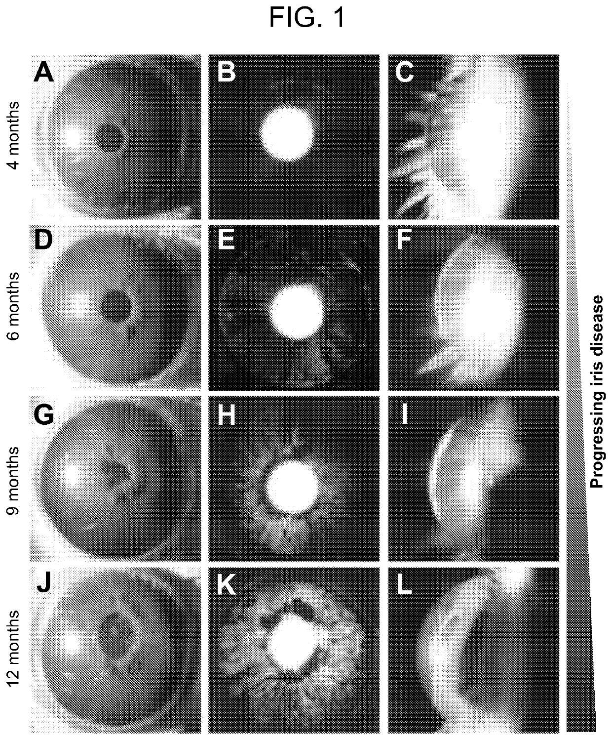
Example 1 DBA / 2J Mice Have a Progressive Iris Disease that Closely Resembles Human Pigmentary Dispersion

[0216]The DBA / 2J (D2) mouse displays many of the hallmark features of human glaucoma. In the D2 mouse, mutations in two genes (GpnmbR150X and Tyrp1b) cause a front-of-the-eye disease that closely resembles human pigmentary dispersion and iris atrophy disease, which cause glaucoma. In the eyes of D2 mice, dispersed iris pigments block channels known as trabecular meshwork in the anterior chamber of the eyes. The trabecular meshwork is responsible for the normal homeostasis of aqueous humor production and outflow. As a consequence of its block...

example 2 progressive

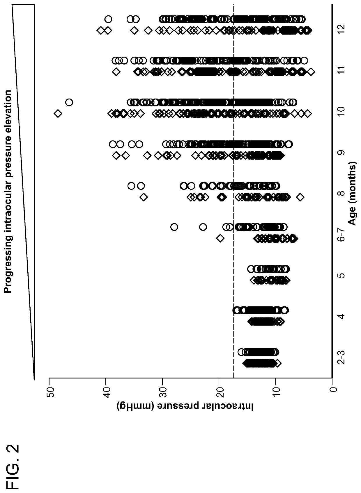
Iris Disease Leads to Increased Intraocular Pressure in DBA / 2J Eyes

[0221]Following the onset of iris disease around 6 months of age, intraocular pressure began to increase (FIG. 2). A typical “normal” IOP in a D2 mouse eye is ˜13 mmHg, which increases progressively from 6 to 12 months, to values that can typically exceed 20 mmHg. By 9 months of age, IOP was / had been elevated in the majority of the eyes of the D2 mice. IOP increases in D2 eyes were age-related and asynchronous, as well as being highly variable. In our colony, females tend to have higher / early onsets of elevated IOP (FIG. 2).

example 3

Eyes of DBA / 2J Mice Have Progressive Visual Dysfunction as Assessed by Pattern Electroretinography (PERG)

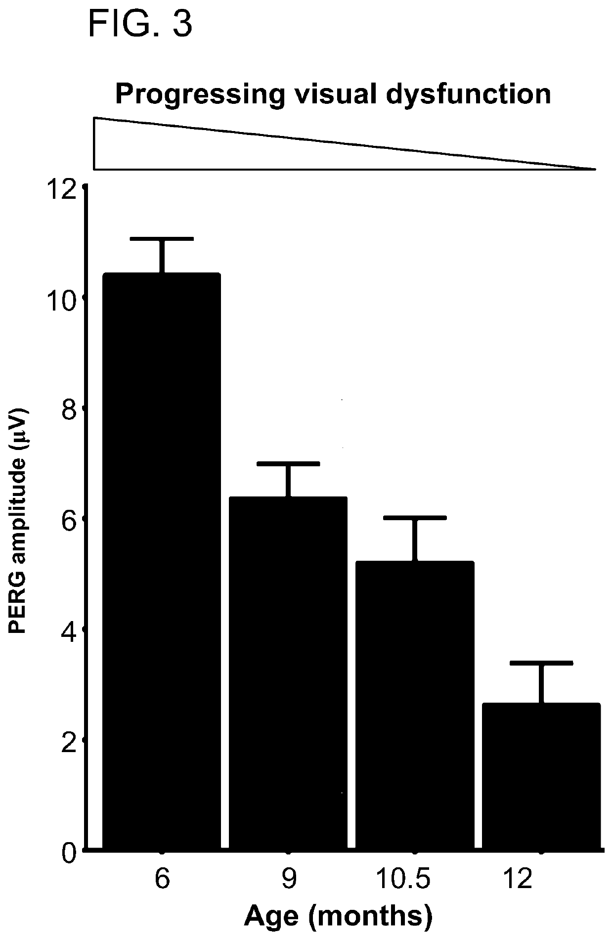
[0222]The retinal ganglion cells (RGCs) are the output neuron of the retina, and their axons make up the optic nerve. Retinal ganglion cells are specifically affected during periods of intraocular pressure that manifests as visual dysfunction prior to gross histological defects, including retinal ganglion cell loss and degeneration of the optic nerve).

[0223]In this example, we used pattern electroretinography (PERG) to measure retinal ganglion cell health in order to detect decreases in visual function prior to degeneration of the optic nerve. PERG assessment of D2 eyes (FIG. 3) demonstrated a progressive loss of visual function that is significant from 9 months of age, which is the time when the majority of the D2 mice eyes had / have had elevated IOP, but is prior to any retinal ganglion cell loss or axon degeneration in the optic nerve. As is shown in FIG. 3, by 12 months of age...


PUM


Abstract

The present invention relates to the use of lipid / fat droplet deposits in retinas of a human subject as a biomarker for diagnosing, prognosing, and / or monitoring retinal neurodegeneration with or without an elevated intraocular pressure in glaucoma.

Description

REFERENCE TO RELATED APPLICATIONS[0001]This application claims the benefit of the filing date under 35 U.S.C. 119(e) of U.S. Provisional Application No. 62 / 429,950, filed on Dec. 5, 2016, the entire content of which is incorporated by reference herein.STATEMENT REGARDING FEDERALLY SPONSORED RESEARCH[0002]The present invention was made with U.S. government support under Grant No. R01 EY011721, awarded by the National Institute of Health (NIH). The U.S. government has certain rights in the invention.BACKGROUND OF THE INVENTION[0003]Glaucoma is an ocular disorder affecting more than 70 million people world-wide and is characterized by cupping of the optic disc due to loss of retinal ganglion cells (RGCs) and the corresponding axons leading to progressive visual loss. It is the leading cause of preventable blindness in the United States. Primary Open Angle Glaucoma (“POAG”) is the most common form of glaucoma.[0004]Several methods are currently available to screen if a human suffers fro...

Claims


Application Information

Patent Timeline
Patent Type & Authority Applications(United States)
IPC IPC(8): A61K49/00A61K9/00A61B3/12A61B3/13A61B3/10
CPCA61B3/13A61K9/0014A61B3/102A61K49/0017A61B3/12A61K9/0019A61K2123/00A61B3/1025
Inventor JOHN, SIMON W.M.WILLIAMS, PETER ALEXANDER
Owner JACKSON LAB THE
Why Eureka
Try Eureka