Fetal ultrasound image processing

a processing and ultrasound technology, applied in image enhancement, ultrasonic/sonic/infrasonic image/data processing, instruments, etc., can solve problems such as difficult tasks and difficult tasks, and achieve the effect of reducing uncertainties or removing them

Active Publication Date: 2020-07-23
KONINKLJIJKE PHILIPS NV
View PDF0 Cites 6 Cited by
  • Summary
  • Abstract
  • Description
  • Claims
  • Application Information

AI Technical Summary

Benefits of technology

[0014]This method enables alignment of a 3D volume image using two reference axes, in order to provide a normalized representation which removes variation resulting from different fetus orientations. A first reference axis is based on the spine, and it may be the direction of the spine in the middle of the spine, or it may be the direction of the vector connecting identified end points of the spine, for example. The spine is a well-defined, unique structure within the fetus that will be readily recognizable to the ultrasound system, particularly when using image recognition techniques.
[0023]The spine is at one edge of the abdomen / thorax, so the vector between the spine and the center of the ellipse (or circle) represents a rotational orientation. In this way, it is possible to normalize the orientation of the 3D volume images based on the torso / abdominal shape of the fetus. As the torso / abdomen is the largest portion of the fetus, it is easily measurable, thereby increasing the accuracy of the alignment.
[0037]In this way, the scale variability between 3D fetal ultrasound images due to gestation age is reduced, and / or the intensity variation due to varying image acquisition gains and imaging conditions may be reduced. The scaling and intensity normalization may take place before or after the reference axis determination and re-orientation.
[0043]In this way, the detection of the spine takes advantages of the strengths of both methods, so that the position of the spine within the 3D fetal ultrasound image can be more robustly determined.
[0055]By generating a classifier for a 3D volume image based on randomly sampled patches of an extracted plane, it is possible to efficiently represent the orientation of the 3D volume image, for example as a binary indicator. The patch size is for example based on a fetal gestation age and a resolution of the 3D fetal ultrasound image.
[0061]This method provides identification of landmarks using the previous machine learning from the testing database, so that the landmarks may be located in an automated way. The 3D orientation processing removes or reduces uncertainties resulting from unknown fetal positions.

Problems solved by technology

This task can be very difficult because of multiple factors, such as the mother morphology, the unknown and highly variable orientation of the fetus as well as well-known US artefacts, in particular the shadowing effect.
However this task is challenging due to the high variability of the fetus orientation at the acquisition time.

Method used


Image

  • Fetal ultrasound image processing
  • Fetal ultrasound image processing
  • Fetal ultrasound image processing

Examples


Embodiment Construction

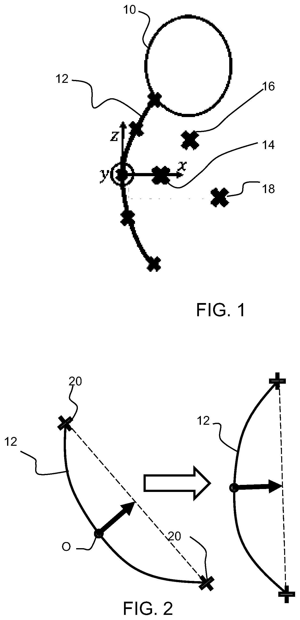
[0083]The invention provides a computer implemented method for processing a 3D fetal ultrasound image. A 3D fetal ultrasound image is obtained (either acquired or received from memory), and the spine is detected within the image. This enables a first reference axis to be defined. A second reference axis is defined perpendicular to the first reference axis, and the 3D fetal ultrasound image is updated (e.g. rotated in 3D space) using the first and second reference axes and an up / down (elevation) orientation detection. This provides a normalization of the orientation of the image, so that a machine learning approach is better able to identify landmarks within new images.

[0084]FIG. 1 shows a representation of a fetus comprising a head 10 and spine 12. The fetus is positioned within 3D space as defined by the axes x,y,z and with an origin at the center of the spine.

[0085]When selecting 2D ultrasound image planes from a 3D ultrasound scan, planes are selected to pass through landmarks of...


PUM


Abstract

A computer implemented method is provided for processing a 3D fetal ultrasound image. A 3D fetal ultrasound image is obtained (either acquired or received from memory), and the spine is detected within the image. This enables a first reference axis to be defined. A second reference axis is defined perpendicular to the first reference axis, and the 3D fetal ultrasound image is updated (e.g. rotated in 3D space) using the first and second reference axes and an up / down (elevation) orientation detection. This provides a normalization of the orientation of the image, so that a machine learning approach is better able to identify landmarks within new images.

Description

FIELD OF THE INVENTION[0001]This invention relates to the processing of fetal ultrasound images.BACKGROUND OF THE INVENTION[0002]In a clinical setting, 2D ultrasound (US) is the preferred scanning protocol for biometry measurements, growth monitoring and anatomy assessment during pregnancy. Obtaining reproducible and accurate values requires strict guidelines to be followed, especially regarding the selection of the standard 2D viewing planes which are used to search for abnormalities or perform biometry measurements such as head and abdomen circumference or femur length. This task can be very difficult because of multiple factors, such as the mother morphology, the unknown and highly variable orientation of the fetus as well as well-known US artefacts, in particular the shadowing effect.[0003]3D US is a more recent imaging technique that has the potential to overcome some of the above mentioned difficulties. In particular, the acquisition of a single 3D volume makes it possible to ...

Claims


Application Information

Patent Timeline
Patent Type & Authority Applications(United States)
IPC IPC(8): G06T7/00A61B8/08G06T7/73
CPCG06T2207/10136G06T7/73G06T2207/30012A61B8/0866G06T2207/20081A61B8/5223G06T2207/30044A61B8/483G06T7/0012G06T3/0093G06T5/006G06T2207/20084
Inventor RAYNAUD, CAROLINE DENISE FRANCOISEROUET, LAURENCECIOFOLO-VEIT, CYBÈLELEFEVRE, THIERRYROUNDHILL, DAVID NIGEL
Owner KONINKLJIJKE PHILIPS NV
Why Eureka
Try Eureka